Table 2-1.
Muscles of the Shoulder
| MUSCLE | ORIGIN | INSERTION | NERVE SUPPLY |
|---|---|---|---|
| Pectoralis major | Medial half of the anterior surface of the clavicle, side and front of the sternum as far as the 6th costal cartilage, front and surfaces of the cartilage of the 2nd through 6th ribs, osseous ends of the 6th and 7th ribs, and aponeurosis of external abdominal oblique | Crest of the greater tubercle of the humerus, lateral lip of the intertubercular groove, deltoid tubercle, and fibrous periosteum of the intertubercular sulcus | Lateral and medial pectoral (C5 and C6 for the clavicular part, and C7, C8, and T1 for the sternocostal part) |
| Pectoralis minor | Aponeurotic slips from the 2nd through 5th ribs, near costal cartilages | Anterior half of the medial border and upper surface of the coracoid process of the scapula | Medial and lateral pectoral (C6, C7, C8) |
| Subclavius | First rib and its cartilage | Inferior surface of the clavicle between the costal and coracoid tuberosities | Nerve to subclavian (C5 or C5 and C6) |
| Deltoid | Lateral border and upper surface of the lateral third of the clavicle, the acromion, and the scapular spine | Deltoid tuberosity of the humerus | Axillary (C5, C6) |
| Supraspinatus | Supraspinous fossa and investing fascia | Shoulder capsule and superior facet of the greater tubercle of the humerus | Suprascapular (C4, C5, C6) |
| Infraspinatus | Infraspinous fossa, scapular spine, investing (deep) fascia, and adjacent aponeurotic septa | Shoulder capsule and middle facet of the greater tubercle of the humerus | Suprascapular (C4, C5, C6) |
| Teres minor | Upper two thirds of the axillary border of the scapula | Shoulder capsule and inferior facet of the greater tubercle of the humerus | Axillary (C4, C5, C6) |
| Subscapularis | Subscapularis fossa | Shoulder capsule and lesser tubercle of humerus and its shaft immediately below the tubercle | Two or three subscapular branches from posterior cord and upper and lower subscapular (C5, C6, C7) |
| Teres major | Inferior angle of the scapula | Medial lip of the intertubercular groove of the humerus | Lower subscapular (C6, C7) |
| Latissimus dorsi | Spine and interspinous ligaments of the lower five or six thoracic vertebrae, upper lumbar vertebrae, thoracodorsal fascia, posterior third of the crest of the ilium, and the lateral surface and upper edge of the lower three or four ribs | Muscle tendon inserts onto the ventral side of the lesser tubercle of the humerus and onto the floor of the intertubercular groove ventral to the tendon of the teres major. The tendon may extend to the greater tubercle of the humerus. | Thoracodorsal (C6, C7, C8) |
Axial
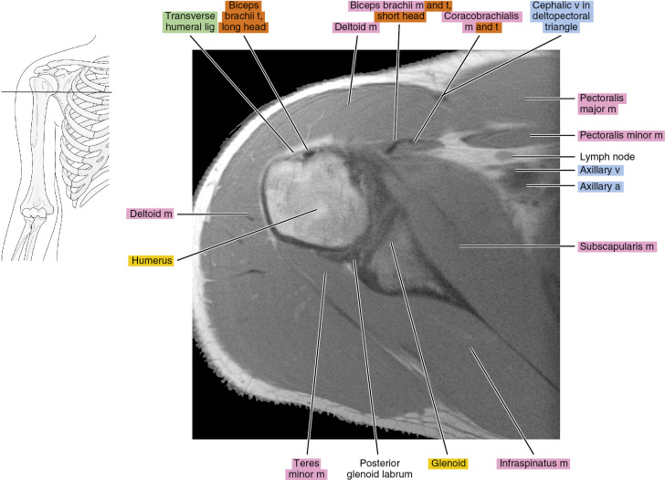
Figure 2.1.1

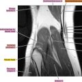
Figure 2.1.2

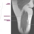
Figure 2.1.3

Figure 2.1.4

Figure 2.1.5

Figure 2.1.6

Figure 2.1.7

Figure 2.1.8

Figure 2.1.9
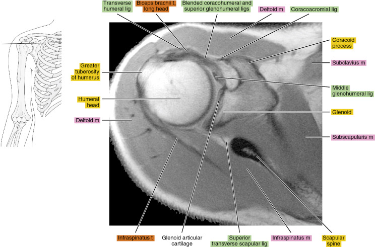
Figure 2.1.10

Figure 2.1.11

Figure 2.1.12

Figure 2.1.13

Figure 2.1.14