Abstract
Multiple pulmonary nodules are most often metastases from a distant primary tumor, but they must be distinguished from a number of inflammatory and infectious diseases that may cause multiple pulmonary nodules or masses. Calcification of nodules is common and most often the result of a healed granuloma from tuberculosis or histoplasmosis. Silicosis and coal worker’s pneumoconiosis both cause upper lobe nodules and masses that may have a characteristic appearance with an upper lobe distribution. Sarcoidosis occasionally causes multifocal opacities that may appear as solid nodules or masses on the chest x-ray, but computed tomography often reveals air bronchograms and confirms the opacities to be consolidations rather than masses. Confirmation of consolidations rather than solid masses makes metastases unlikely.
Keywords
coal worker’s pneumoconiosis, granulomas, masses, metastases, sarcoidosis, silicosis
Questions
- 1.
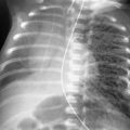
A 50-year-old man admitted with a history of weight loss had the examinations shown in Fig. 21.1, A and B . Which one of the following procedures would most likely determine the site of a primary tumor?
- a.
Physical examination of the testis.
- b.
Total spine radiograph.
- c.
Radionuclide bone scan.
- d.
Computed tomography (CT) scan of the brain.
- e.
Magnetic resonance imaging (MRI) of the knee.

Fig. 21.1
- a.
- 2.
Which one of the following primary tumors is least likely to metastasize to the lung?
- a.
Melanoma.
- b.
Osteosarcoma.
- c.
Astrocytoma.
- d.
Adenocarcinoma of the colon.
- e.
Choriocarcinoma.
- a.
- 3.
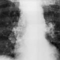
Which one of the following diagnoses is most likely in the patient shown in Fig. 21.2, A and B ?
- a.
Tuberculosis.
- b.
Primary lung cancer.
- c.
Metastases.
- d.
Coal worker’s pneumoconiosis.
- e.
Histoplasmosis.

Fig. 21.2
- a.
Discussion
One of the first decisions in the evaluation of the chest radiograph showing multiple pulmonary opacities is to distinguish multiple nodules and masses from areas of pulmonary consolidation. Nodules and masses are more circumscribed and homogenous, whereas consolidating lesions have less circumscribed borders and are less solid, often containing aerated alveoli and air bronchograms. Loss of definition of the border implies that the process is spreading into the lung parenchyma, either following interstitial planes or actually spilling into the alveolar spaces. Loss of definition is therefore more consistent with a consolidating process. High-resolution CT has been used to better characterize the borders of a variety of metastatic nodules and has shown that metastases may be locally invasive, 247 which would account for the phenomenon of nodules growing and becoming less circumscribed.
A second major problem confronting the radiologist in the evaluation of the patient with multiple pulmonary nodules is the perception of the nodules. Small, peripheral pulmonary nodules are often not detectable on the chest radiograph. These nodules are obscured by surrounding vascular opacities or may be incompletely surrounded by aerated lung. They are also obscured by overlying ribs. CT reliably detects peripheral subpleural nodules and nodules obscured by the heart, mediastinum, and vessels. 399 , 624 Spiral CT with a single breath-hold technique reduces artifacts and has reduced the risk of missing small nodules that change position with respiratory motion. CT is the most sensitive procedure available for detecting pulmonary nodules. By increasing the sensitivity of the procedure, it has become the optimal technique for staging a variety of cancers; however, as the sensitivity increases, the specificity decreases. Although CT detects more metastatic nodules than chest radiography, it also detects more benign nodules caused by granulomas and unrelated scars. The decision to perform a CT scan should be based on the primary diagnosis and the plan of therapy. When the radiographs show multiple, bilateral pulmonary nodules, the value of detecting an additional number of nodules depends on the mode of therapy planned for the patient. If the therapy will not be influenced by the detection of additional nodules, it is doubtful whether the more expensive and complicated procedure is justified. When resection of a metastatic nodule is contemplated, the detection of additional nodules will change the treatment plan, and the preoperative CT scan is essential.
Neoplasms
From the preceding discussion, it should be apparent that metastatic disease is the most important cause of multiple pulmonary nodules in today’s practice. The list of primary tumors that metastasize to the lung is long ( Chart 21.1 ). 197 , 361 In most cases, multiple pulmonary metastases are detected after the primary lesion, thus making the presumptive diagnosis of metastases from the known primary lesion a very secure diagnosis ( Fig. 21.3 ). Although this is a reliable assumption for larger masses that are detected by chest radiograph, very small occult opacities detected by CT may often represent benign nodules. These very small opacities must be cautiously evaluated when their detection may influence the choice of therapy, sometimes even justifying biopsy. Multiple pulmonary metastases constitute a relatively unusual presenting complaint that is sometimes followed by an extensive search for an occult primary tumor. One primary tumor that is well known for this presentation is testicular carcinoma. When a male patient’s chest radiograph shows multiple pulmonary nodules and masses of varying size, testicular carcinoma should be one of the first considerations. Physical examination may confirm the diagnosis (answer to question1 is a ), but detection of very small occult tumors often requires testicular ultrasound. Other primary tumors that may be occult but metastasize to the lungs include melanoma, ovarian carcinoma, breast cancer, renal cell carcinoma, colon cancer, and gastrointestinal tumors. Most other tumors that have a high rate of pulmonary metastases have local findings and are less likely to present as an occult primary. Spontaneous pneumothorax and multiple pulmonary nodules together are an unusual combination that is nearly diagnostic of osteosarcoma ( Fig. 21.4 ), although it has been encountered with other tumors (e.g., Wilms tumor). This combination should suggest the diagnosis of osteosarcoma, but primary bone tumors are rarely occult. They usually cause pain and are diagnosed via radiography and combinations of CT, MRI, and radionuclide bone scans. Patients with primary bone tumors are more likely to have occult pulmonary metastases at the time of diagnosis. The unsuspected metastases may be diagnosed by radiography or may require CT.
- I.
Neoplastic
- A.
Malignant
- 1.
Metastases (kidney, gastrointestinal tract, uterus, ovary, testes; melanoma, sarcoma)
- 2.
- 3.
Posttransplantation lymphoproliferative disorder 119 , 224 , 474
- 4.
Kaposi sarcoma (acquired immunodeficiency syndrome [AIDS] related) 126
- 1.
- B.
Benign
- 1.
Hamartoma 510
- 2.
Arteriovenous malformation 135 or hemangioma
- 3.
- 4.
Benign metastasizing leiomyoma 7
- 1.
- A.
- II.
Inflammatory
- A.
Fungal 371
- B.
- C.
Tuberculosis 388 (typical and atypical)
- D.
Parasites
- E.
Atypical measles 661
- F.
Inflammatory pseudotumors 10 (e.g., fibrous histiocytoma, plasma cell granuloma, 283 hyalinizing pulmonary nodules)
- G.
Q fever 385
- H.
Sarcoidosis
- A.
- III.
Vascular
- 1.
Rheumatoid nodules and Caplan syndrome 59 , 283
- 2.
Granulomatosis with polyangitis 4
- 3.
- 4.
Septic emboli
- 1.
- IV.
Posttraumatic (organizing hematoma) 499
- V.
Chronic renal failure (calcified nodules) 77
Related posts:


Stay updated, free articles. Join our Telegram channel

Full access? Get Clinical Tree


