• Ruxolitinib is an oral J AK1 and JAK2 inhibitor approved by FDA for treatment of intermediate or high-risk myelofibrosis
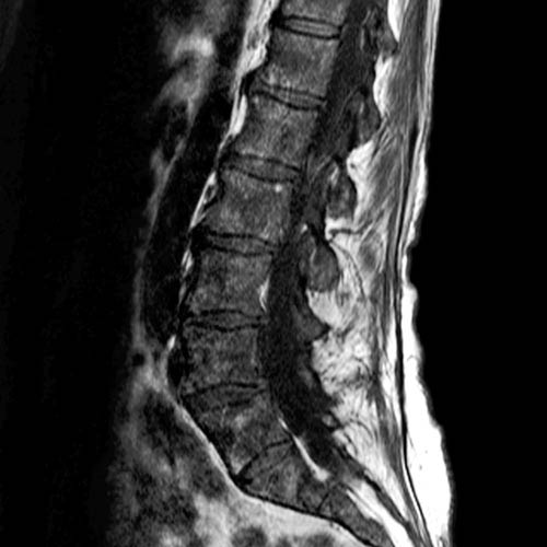
(Left) Sagittal T1WI MR in a 70-year-old woman with a long history of thrombocytosis and subsequent development of pancytopenia shows extensive, mildly heterogeneous low signal intensity in bone marrow. Biopsy confirmed myelofibrosis.
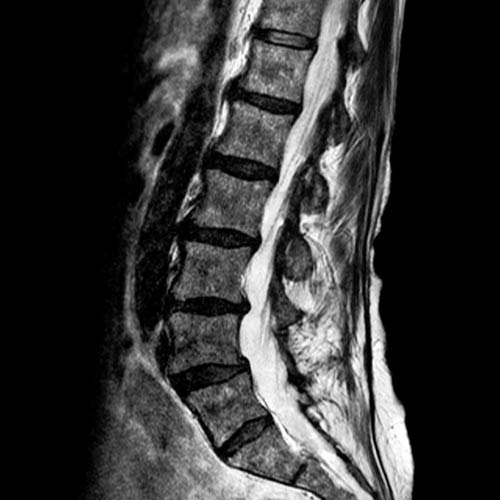
(Right) Sagittal T2WI MR in the same patient shows that bone marrow is low signal intensity. The patient was refractory to treatment, but she survived 8 years from diagnosis of myelofibrosis.
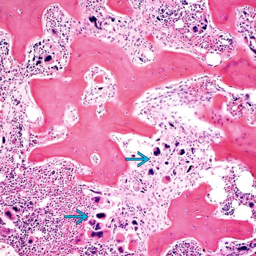
(Left) Marked osteosclerosis, myelofibrosis, and megakaryocytic pleomorphism are the hallmark histologic features of primary myelofibrosis. Megakaryocytes range from small to large and are hyperchromatic and clustered .
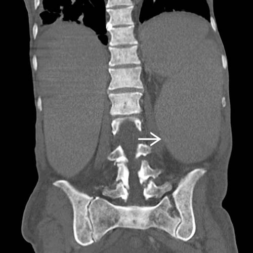
(Right) Coronal bone CT in the same patient shows bone sclerosis and splenomegaly . Extramedullary hematopoiesis most commonly involves the spleen, but it may also be seen in paraspinous soft tissues.
TERMINOLOGY
Synonyms
• Myelosclerosis
• Agnogenic myeloid metaplasia
Definitions
• Myelodysplastic syndrome characterized by marrow replacement with fibrous tissue
IMAGING
Radiographic Findings
• Radiography
Usually normal; bone sometimes diffusely sclerotic
Extramedullary hematopoiesis
– Splenomegaly, hepatomegaly
Only gold members can continue reading. Log In or Register to continue


 .
.
 . Extramedullary hematopoiesis most commonly involves the spleen, but it may also be seen in paraspinous soft tissues.
 Extramedullary hematopoiesis


