Nasopharynx
The roof of the nasopharynx is formed by the sphenoid sinus and upper clivus. The posterior margin is formed by the lower clivus and upper cervical spine. Anteriorly lies the nasal cavity. The lateral walls are formed by the pterygoid plates (anteriorly) and the fascia and muscles of the airway (posteriorly). The nasopharynx is separated from the oropharynx below by the soft palate. The levator and tensor veli palatini muscles arise from the skull base, attach to the soft palate, and function to elevate and tense the palate. The pharyngobasilar fascia holds the airway patent. It surrounds the mucosa, superior constrictor, and levator veli palatini muscles, and separates the nasopharyngeal mucosal space from surrounding fascial spaces.
The nasopharyngeal mucosal space contains mucosa, adenoidal tissue, the superior constrictor muscle, the torus tubarius, levator veli palatini muscles, and many minor salivary glands. The mucosa and normal adenoidal tissue are high signal intensity on T2-weighted MR images. The eustachian tubes end in the torus tubarius (a lateral cartilaginous enlargement). The lateral pharyngeal recess (fossa of Rosenmüller) is formed by mucosal reflection over the longus colli and capitis muscles (flexors of the cervical spine), and is the site of origin for the vast majority of nasopharyngeal carcinomas (NPC). The levator veli palatini muscle is intrapharyngeal, just lateral to the torus tubarius. The tensor veli palatini muscle is lateral to the pharyngobasilar fascia and surrounded by fat. The retropharyngeal space is a potential space between the pharyngeal constrictor muscles anteriorly and the prevertebral muscles posteriorly. The prevertebral (perivertebral) space is bounded by the prevertebral fascia anteriorly and the vertebral bodies posteriorly, contains the prevertebral muscles, and extends from the skull base to the coccyx.
Infection involving the nasopharynx is usually secondary to either dental or tonsillar infection. Dental infection can spread to the masticator and prestyloid parapharyngeal spaces, in addition to causing osteomyelitis. Tonsillar infection can result in an abscess that can extend to the retropharyngeal space or the prestyloid parapharyngeal space.
Benign, incidental soft tissue masses in the nasopharynx include adenoidal hypertrophy (seen in children and young adults), and the Tornwaldt cyst. The latter is very common, seen in 4% of patients, and is a small midline cyst that lies along the posterior nasopharyngeal wall. A Tornwaldt cyst will have high signal intensity on T2, reflecting fluid, and intermediate to high signal intensity on T1-weighted images, dependent on the protein concentration.
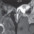
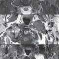
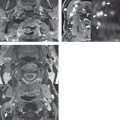
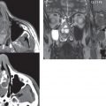
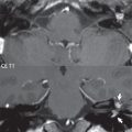
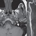
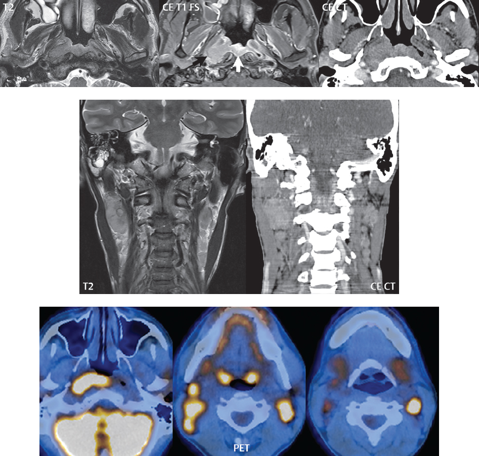
Nasopharyngeal carcinoma is by far the most common malignant tumor of the nasopharynx, strongly associated with Epstein-Barr virus infection. The lateral pharyngeal recess is the most common site of origin. These tumors often cause eustachian tube obstruction, due to involvement of the levator veli palatine muscle, resulting in a middle ear effusion (Fig. 2.8). A tumor in this location can grow in any direction, with lateral extension most common. Eighty to 90% of patients have nodal involvement on presentation (retropharyngeal, level II and level V) ( Fig. 2.85 ). Involvement of lymph nodes can be suggested on many different bases—by size criteria, with visualization of necrosis, and by restricted diffusion, with FDG PET extremely valuable for identification of tumor involvement (above a certain size). It is important to note that radiation therapy causes a substantial change in appearance of tissues in this region on both CT and MR, with fibrosis having low signal intensity on both T1- and T2-weighted images.
Rhabdomyosarcoma is the most common sarcoma in pediatrics and young adults. Forty percent of these lesions occur in the head and neck, with the most common locations being the orbit and nasopharynx. This tumor is locally invasive, often with bone destruction and perineural tumor extension.
Stay updated, free articles. Join our Telegram channel

Full access? Get Clinical Tree


