Abstract
Teratomas are the most common histologic type of fetal tumors. The neck is the most common location after the sacrococcygeal area. A neck teratoma may be associated with neonatal mortality in 80% to 100% of cases if delivery is not managed properly. Prenatal diagnosis, ex utero intrapartum treatment (EXIT), and intensive neonatal care are essential to improve the management. EXIT allows adequate respiratory assistance and improves neonatal survival in cases of tracheal obstruction. Early resection is the treatment of choice.
Keywords
fetal tumors, neck masses, cervical teratoma, congenital airway obstruction, ex utero intrapartum treatment (EXIT)
Introduction
Fetal tumors are rare; teratomas are the most common histologic type. The neck is the most common location after the sacrococcygeal area for teratomas. The tissues found in fetal and infant teratomas are essentially the same regardless of the site of origin. A neck teratoma may be associated with neonatal mortality in 80% to 100% of cases if delivery is not managed properly. For large masses, adequate prenatal diagnosis allowing planned delivery with ex utero intrapartum treatment (EXIT) and intensive neonatal care are essential to improve the management.
Disease
Definition
A normally benign tumor in the neck composed of multiple tissues derived from all three germ layers of the embryonic disk—ectoderm, mesoderm, and endoderm—with a high potential of growing in excess.
Prevalence and Epidemiology
The prevalence of fetal tumors is difficult to establish; however, it is estimated to be from 1.7 : 100,000 to 13.5 : 100,000 live births. Cervical teratomas are found in about 1 : 20,000 to 1 : 40,000 live births, accounting for 3% to 6% of all neonatal teratomas.
The presentation is sporadic, without an apparent relationship to race, maternal age, parity, or fetal sex. Some cases of familial recurrence have been reported.
Etiology and Pathophysiology
The development of fetal tumors does not match the same processes as tumors observed in adults. In fetuses, tumors may result from failure of developing tissues to undergo normal cytodifferentiation and maturation.
Cervical teratomas may originate from the palate, nasopharynx, or thyrocervical area. They are usually closely related to, but do not arise from, the thyroid gland. Mature or immature neuroglial tissues are the most frequent component, but cartilage, respiratory epithelium, and ependyma-lined cysts are common. Malignancy is extremely rare. Immature elements present do not express the biologic behavior.
Manifestations of Disease
Clinical Presentation
Ultrasound (US) diagnosis is usually made at routine anatomic scan. Polyhydramnios, caused by esophageal compression, is present in 30% to 40% of cases. More rarely, the diagnosis is established after an episode of preterm labor caused by polyhydramnios.
Imaging Technique and Findings
Ultrasound.
Prenatal US diagnosis of a teratoma can be made as early as the first trimester, but they are usually detected on routine second-trimester screening or after an initial diagnosis of polyhydramnios. The key sign is the distortion in neck contour by the presence of an asymmetric, unilateral, and well-encapsulated mass ( Fig. 71.1 ; ). The tumor is usually large and bulky, with mixed echostructure of cystic and solid components. Echogenic foci of calcifications, present in about half of all cases, are virtually pathognomonic of teratoma ( Fig. 71.2 ). Located anterior to the neck, the tumor may produce a mass effect on surrounding tissues from the ear to the jaw or extend into the mediastinum ( Fig. 71.3 ; ). Intracranial and orbital extensions are rare. Large tumors result in severe hyperextension of the fetal head ( ).


Polyhydramnios indicates severity, since it reflects impaired swallowing by mouth obstruction or esophageal compression ( Fig. 71.4 ).

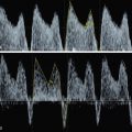
Solid parts are often very vascular and with arteriovenous shunts. Color Doppler shows the intensity and characteristics of the tumor vascularization ( Fig. 71.5 ). Three-dimensional US may provide additional detailed information on location, extension, and intracranial spread ( Figs. 71.6 and 71.7 ).