Fig. 1
Serous cystadenoma. (a) Surgical specimen (pancreaticoduodenectomy): serous cystadenoma with typical microcystic appearance and central scar (white arrow) with small calcification (black arrow). (b) Surgical specimen (pancreaticoduodenectomy): serous cystadenoma with typical microcystic appearance and eccentric scar (arrow). (c) Surgical specimen (pancreaticoduodenectomy): serous cystadenoma with typical microcystic appearance and very large central scar (arrow). (d) Surgical specimen (distal pancreatectomy): serous cystadenoma with typical microcystic appearance with no macroscopically evident scar. (e) Histopathology: serous cystadenoma with typical single layer of clear cuboidal epithelial cells lining the cysts with typical subepithelial vessels (asterisks)
Fig. 2
Oligocystic serous cystadenoma. (a, b) Surgical specimen (distal pancreatectomy): pancreatic neoplasm (a) in the pancreatic body, presenting as a lobulated (arrow in a) cystic mass delimitated by a very thin wall. At cut section (b), the lesion is oligocystic. The Wirsung duct (W in b ) is slightly dislocated, but normal in caliber. (c) Surgical specimen (intermediate pancreatectomy): oligocystic serous cystadenoma with typical very thin septa (arrow)
Fig. 3
Serous cystic neoplasm. (a–c) Ultrasound study: pancreatic head lobulated cystic lesion (arrow in a) with tiny vessels at Doppler (a, b), along the septa, resulting centrally orientated and vascularized (arrow in c) at CEUS (c)
Fig. 4
Serous cystic neoplasm. (a, b) Ultrasound study: pancreatic head lobulated cystic lesion (arrow in a) with tiny vascularized septa (arrow in b) at CEUS (b) resulting centrally orientated. (c) MRI: the cystic lesion is typical, hyperintense (arrow) on T2-weighted images. (d ) Surgical specimen (pancreaticoduodenectomy): oligocystic serous cystadenoma with central scar (Courtesy of Piccin Editore, Milan, Italy)
Fig. 5
Serous cystic neoplasm. (a, b) CT study: serous microcystic neoplasm, presenting as a hypodense mass in the baseline (a) and dynamic (b) scans, with central calcification (arrow in a). (c, d) CT study: pancreatic head serous oligocystic neoplasm presenting as a cystic hypodense mass in the baseline (arrow in c) and dynamic (arrow d) scans
Fig. 6
Serous cystadenoma. (a–c) CT study: serous microcystic neoplasm (arrow in a), better visible on coronal (b) and minimum-intensity projection (c) reconstructions, with typical lobulated contours (arrow in c)
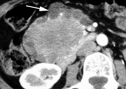
Fig. 7
Big serous cystic neoplasm. Dynamic CT: pancreatic head big serous cystadenoma with prevalent central microcystic appearance, resulting hypodense, and with relatively larger cystic portions, typically located at the periphery (arrow) of the mass
Fig. 8
Small serous cystic neoplasm. (a) Ultrasound study: small lobulated microcystic lesion (calipers) of the pancreatic body. (b) CT: small lobulated hypodense lesion (arrow) of the pancreatic body. (c) MRI: small lobulated microcystic lesion (arrow) of the pancreatic body
Fig. 9
Serous cystadenoma. (a–d) MRI study: small microcystic lesion of the uncinate process of the pancreas, appearing hypointense on T1-weighted fat-saturated (a) images and hyperintense on T2-weighted (b) images. The lesion shows typically lobulated contours (arrow in b). On MRCP (c), the absence of communication between the microcystic lesion (arrow in c) and the Wirsung duct (W in c) is perfectly documented. In the dynamic phase (d), the lesion shows microcystic aspect with vascularized thin septa and lobulated contours (arrow in d)
Fig. 10
Lobulated contour of serous cystic neoplasm. (a, b) MRI study: oligocystic serous cystadenoma with typical lobulated contours (arrow in a) in the body of the pancreas. On MRCP (b), the typical morphology of the cystic lesion with lobulated contours is visible. The Wirsung duct (W in b) is normal and faintly visible through the cystic lesion as an indirect sign of separation between the two structures
Fig. 11
Serous cystadenoma. (a, b) MRI study: microcystic mass, hyperintense on T2-weighted images with small central hypointense scar (arrow in a). The multiple microcystic lesion septa and also the fibrovascular small scar are well vascularized in the dynamic phase (b). (c) Surgical specimen (distal pancreatectomy) of different case: microcystic serous cystadenoma with central scar (arrow)
Fig. 12
Serous cystadenoma. (a–g) MRI study: microcystic mass, hyperintense on T2-weighted axial (a) and coronal (b) images, with big central hypointense scar (arrow in b). At DWI, the cystic lesion shows drop of signal intensity moving from low (c) to high (d) b value. In the dynamic pancreatic (e), venous (f), and late (g) phases, the lesion is well vascularized with progressive accumulation of contrast medium in the scar, resulting less visible (arrow in g) in the late phase. (h) Surgical specimen (distal pancreatectomy): microcystic serous cystadenoma with big central scar (arrow)
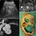
Fig. 13
Serous cystic neoplasm. (a) Ultrasound study: microcystic serous cystadenoma of the pancreatic body, appearing mainly hyperechoic (arrow), with the larger cystic portion (asterisk) typically located at the periphery of the lesion. (b) MRI study: microcystic appearance of the mass on T2-weighted images with central scar and larger cystic portion (asterisk) typically located at the periphery of the lesion. (c, d) Surgical specimen (distal pancreatectomy) of different case: cystic mass with larger cyst (arrow in c) bulging the inferior profile of the pancreas. The cut section confirms a mixed microcystic–oligocystic serous cystadenoma with larger cyst (asterisk in d) in the periphery of the mass
Fig. 14
Huge serous cystadenoma. (a, b) MRI study: huge microcystic mass, hyperintense on T2-weighted images (a) with central scar and evident septa vascularization in the dynamic phase (b). (c, d) Surgical specimen (distal pancreatectomy) of different case: huge cystic mass (M in c) involving completely the body and tail of the pancreas with typical microcystic aspect on the cut section (d) in which the central scar is evident
Fig. 15


Massive serous cystadenoma. (a–d) MRI study: massive microcystic mass hyperintense on coronal T2-weighted images (a, b) with central scar (arrow in a) and normal common bile duct (C in b). MRCP (c) highlights the massive dimensions of the serous cystadenoma, confirming a common bile duct (arrow in c) regular in caliber. At dynamic MRI (d), multiple intralesional extremely thin septa enhancement is visible
Related posts:


Stay updated, free articles. Join our Telegram channel
Full access? Get Clinical Tree
