Fig. 8.1
(60 s uptake image, posterior view, LPO view, and RPO view): both kidneys show good uptake of radiotracer, with homogeneous intraparenchymal distribution; no cortical defects are evident in both kidneys
8.1.2.2 DMSA Renal Scan in Acute Pyelonephritis
DMSA renal scan in a 4-month-old baby, with diagnosis of acute pyelonephritis. No evidence of pathological findings was detected by ultrasonography, while DMSA scan showed a characteristic acute pyelonephritis pattern. Cystography performed after 1 month revealed bilateral high-grade vesicoureteral reflux (VUR). DMSA renal scan in acute pyelonephritis allows an early detection of kidney damage and is characterized by a high prognostic value in identifying children with higher risk of renal impairment (Fig. 8.2a, b).
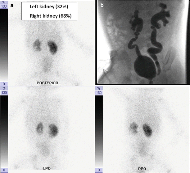
Fig. 8.2
(a) DMSA renal scan (posterior view, LPO view, and RPO view): left kidney is smaller than the other one, with global reduction of radiotracer uptake (acute flogosis); two cortical defects are evident, one in the upper pole and the larger one in the lower pole, respectively; right kidney shows good uptake of radiotracer with decreased uptake in upper and lower poles, without cortical defects. A very low target/background ratio is also evident. (b) Cystography: bilateral high-grade vesicoureteral reflux (Grade IV)
8.1.2.3 DMSA Renal Scan in Follow-up ofVUR: Evaluation of Stable Scars
DMSA renal scan in an 8-year-old child with renovascular hypertension and treated bilateral VUR (Fig. 8.3).
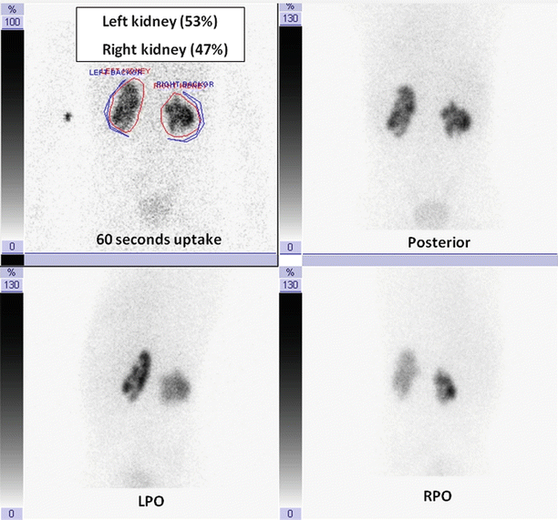
Fig. 8.3
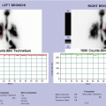
(60 s uptake image, posterior view, LPO view, and RPO view): radiotracer uptake is reduced in both kidneys with bilateral multiple cortical defects, in particular, in the right one. A low target/background ratio is also evident
8.1.2.4 DMSA Renal Scan in Diagnosis of Renal Scars in High-Grade VUR
Severity of VUR is not always proportional to kidney damage, as it is showed in the following cases (Fig. 8.4).
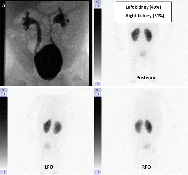


Fig. 8.4
(a) No evidence of scars in relation to high-grade VUR. DMSA renal scan (posterior view, LPO view, and RPO view): both kidneys show good uptake of radiotracer, with homogeneous intraparenchymal distribution; no cortical defects are evident in both kidneys. (b) Evidence of a small scar in relation to high-grade VUR. DMSA renal scan (posterior view, LPO view, and RPO view): left kidney is smaller than the other one, with globally reduced radiotracer uptake and a central area devoid of tracer corresponding to dilated renal pelvis; a small focal cortical defect is evident in the upper pole. Right kidney shows good uptake of radiotracer with homogeneous intraparenchymal distribution and no evidence of cortical defects. (c) Evidence of multiple bilateral scars. DMSA renal scan (posterior view, LPO view, and RPO view): both kidneys are small with irregular shape; radiotracer intraparenchymal distribution is nonhomogeneous, and multiple cortical defects are evident in both kidneys, in particular, in the left one. A very low target/background ratio is also evident.
8.1.2.5 DMSA Renal Scan in Assessment of Renal Function and in Diagnosis of Cortical Defects in Posterior Urethral Valves
A 6–month-old baby, with chronic renal failure secondary to posterior urethral valves.
- (a)
Cystography: bilateral high-grade vesicoureteral reflux (V grade on the left, IV grade on the right) with dilated proximal urethra and radiological signs of posterior urethral valves.
- (b)
DMSA renal scan (posterior view, LPO view): no visualization of right kidney; left kidney has got an irregular shape and shows globally reduced uptake of radiotracer with nonhomogeneous intraparenchymal distribution. A very low target/background ratio is also evident.
8.1.2.6 DMSA Renal Scan in Obstructive Uropathy (With or Without Scars)
- (a)
DMSA renal scan (posterior view) in left hydronephrosis with no evidence of cortical defects. Left kidney is very much enlarged, with mildly irregular shape; it shows nonhomogeneous intraparenchymal distribution of radiotracer, with the evidence of a large area dilated renal pelvis; renal parenchyma is thin, with no cortical defects. Radiourine is evident in dilated pelvis. Right kidney shows good uptake of radiotracer, with homogeneous intraparenchymal distribution and no evidence of cortical defects (left DRF: 55 %; right DRF: 45 %).
- (b)
DMSA renal scan (posterior view) in right hydroureteronephrosis with no evidence of cortical defects. Left kidney shows good uptake of radiotracer, with homogeneous intraparenchymal distribution. Right kidney shows good uptake of radiotracer with nonhomogeneous intraparenchymal distribution for the presence of an area devoid of tracer corresponding to dilated renal pelvis; no cortical defects are evident in both kidneys (left DRF: 48 %; right DRF: 52 %).
- (c)
DMSA renal scan (posterior view) in right kidney hydronephrosis with renal impairment and multiple cortical defects. Left kidney shows good uptake of radiotracer, with homogeneous intraparenchymal distribution and no evidence of cortical defects. Right kidney shows reduced uptake of radiotracer, with the evidence of a large area devoid of tracer corresponding to dilated renal pelvis and collecting system; renal parenchyma is thin, and multiple cortical defects are evident (left DRF: 71 %; right DRF: 29 %).
- (d)
DMSA renal scan (posterior view) in staghorn calculi of left kidney with low renal function. Left kidney is smaller than the other one, with irregular shape, and shows severe reduction of radiotracer uptake. Intraparenchymal distribution is not homogeneous, and many areas devoid of tracer (corresponding to renal litiasis) are evident with multiple focal cortical defects. Right kidney shows good uptake of radiotracer, with homogeneous intraparenchymal distribution and no evidence of cortical defects (left DRF: 28 %; right DRF: 72 %).
Related posts:


Stay updated, free articles. Join our Telegram channel

Full access? Get Clinical Tree


