Fig. 6.1
(a, b) The qualitative analysis of basic dynamic study reveals an intense concentration of tracer in the renal parenchyma and rapid washout of both kidneys (a). Renogram curves confirm qualitative interpretation of the study, showing normal tracer uptake in both kidneys, bilateral regular transit with rapid urinary excretion; the basic renogram also provides a symmetrical measurement of relative renal function (b). IRC images show a complete bladder voiding in the absence of VUR (c). Time-activity curves of bladder and kidneys confirm a regular and complete bladder voiding without radioactivity increment corresponding to kidneys and ureters (d)
Case 6.3 Asymptomatic Residual Monolateral Reflux (Grade II) After Endoscopic Correction of Bilateral Vesicoureteral Reflux
A 2-year-old girl referred to our center for recurrent pyelonephritis. An IRC showed a bilateral vesicoureteral reflux (Grade II); thus, we performed an endoscopic treatment of VUR, followed by antibiotic prophylaxis for 1 month. During follow-up, the child has presented urinary tract infection no longer, but an IRC performed after about 1 year of treatment, as control treatment effectiveness, documented persistence of left-sided vesicoureteral reflux (Fig. 6.2c, d). Therefore, the child underwent ureteral reimplantation, and currently she is asymptomatic.
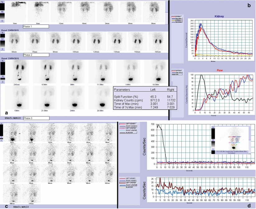
Fig. 6.2
(a, b) A 3-year-old child with a urinary tract infection episode. Having acquired the control of urination, a renal scintigraphy with IRC was performed. The scintigraphic study showed a left kidney slightly smaller than the contralateral one with a normal renal function and a low-grade reflux on the left side. As the child had a unilateral low-grade VUR without nephropathy, he was followed on surveillance by periodic urinalysis and ultrasonography scans, and he is currently asymptomatic. In general, in fact, low-grade refluxes (Grades I–II) have high rates of spontaneous resolution (usually more than 80 %) in young children. By the qualitative assessment of scintigraphic images, both kidneys are located in their own proper sides with regular perfusion. Left kidney is slightly smaller than the contralateral one, even if both kidneys show normal morphology, renal uptake, and urinary excretion. (a) Both renogram curves show normal slope of parenchymal phase and regular elimination of tracer from the kidneys (left curve is slightly less wide in relation to the size of the kidney). The split renal function is sufficiently symmetric (b). (c, d) During bladder voiding (regular and complete), a synchronous radiourine reflux limited to left ureter is observed (Grade I) (c). This scintigraphic finding is confirmed by time-activity curve of the left ureter without radioactivity increment corresponding to homolateral kidney (d)
Case 6.4 Persistent Urinary Tract Infections Secondary to Residual Bilateral Reflux (Grade II–III) After Endoscopic Correction in Bilateral Duplex Collecting System
A 1-year-old baby with right high-grade vesicoureteral reflux in double system. She was treated with antibiotics for several episodes of pyelonephritis in the first months of life. During follow-up, a DMSA scan showed double bilateral system with good function of both kidneys, even if renal scars were bilaterally present in lower systems. The child continued to present febrile urinary infection after endoscopic treatment of vesicoureteral reflux, and a cystography showed persistence of VUR (in both systems of left kidney and in the upper right system). Therefore, a new endoscopic treatment of reflux was performed at 18 months of life, but next IRC showed persistence of bilateral vesicoureteral reflux. Due to the close proximity of the ureteral orifices, endoscopic correction of vesicoureteral reflux in duplicated collecting systems is challenging to be effective, avoiding secondary obstructive megaureter.
Case 6.5 Relapse of High-Grade Vesicoureteral Reflux (Grade II–III) After Acquisition of Bladder-Voiding Control in Child with Reflux Nephropathy and Retensionist Habit
A 3-year-old child with previous recurrent pyelonephritis, followed for bilateral high-grade VUR (treated endoscopically at 18 months) and bilateral nephropathy (at 2 months of life, a DMSA scan showed bilateral renal scarring with greater impairment of the right kidney).
After acquisition of bladder-voiding control, the child had a retentionist attitude, and he had an acute pyelonephritis, despite a time-urination management. An IRC was performed revealing a bilateral vesicoureteral reflux (obtained with high bladder filling), with a severe reflux nephropathy at the right side. VUR highlighted by the scintigraphic study was confirmed by a subsequent cystography, and patient underwent ureter reimplantation (Figs. 6.3, 6.4, and 6.5).
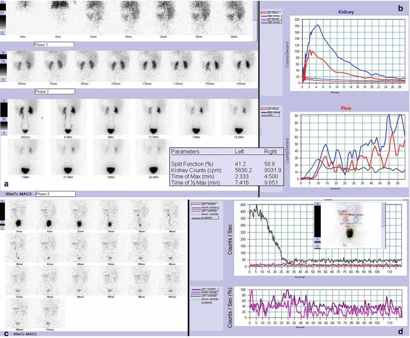
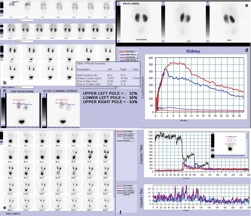
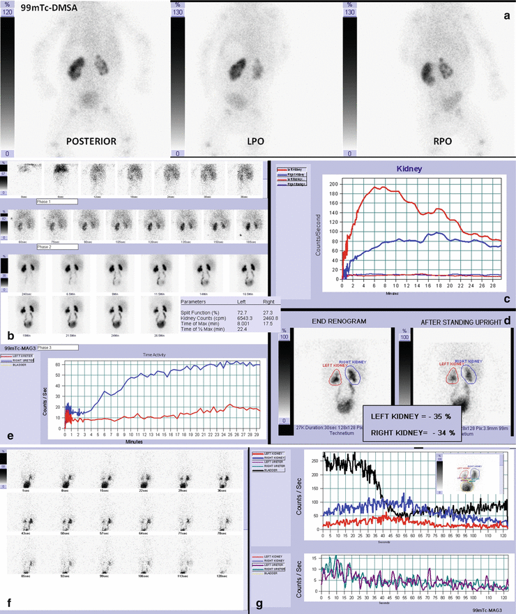
Fig. 6.3
By qualitative assessment of scintigraphic images, both kidneys are located in their own proper sides with synchronous but asymmetrical perfusion (reduced on the left side). Left kidney is small with a slightly irregular morphology; left renal uptake is preserved, and urinary excretion is normal. Right kidney shows an intense concentration of tracer in the renal parenchyma and a rapid washout (a). Left renogram curve (less wide than the contralateral one) shows preserved slope of parenchymal phase and regular elimination of tracer. The shape of the right renogram curve is normal in all phases. The split renal function is slightly asymmetric (relatively reduced on the left) (b). (c, d) IRC images show a complete and regular bladder voiding. During bladder voiding, a synchronous radiourine reflux is present on the left side that reaches the renal pelvis without dilatation (Grade II) (c). Time-activity curves of bladder and kidneys reveal a regular and complete bladder voiding, with a radioactivity increment corresponding to left ureter and kidney (d)
Fig. 6.4
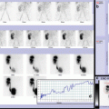
(a) At 8 months of life, a DMSA scan shows a double bilateral system with good uptake of the radiopharmaceutical in both kidneys; tracer concentration is regular in the upper systems, while a nonhomogeneous distribution is detected in the lower systems due to focal cortical defects (more marked scars on the left). No evidence of parenchymal defects is observed in the upper systems.(b, c) At 26 months of life, a MAG3 renal scan is performed showing a good function of the right kidney (both systems) and a double left kidney district with hypofunction and scars of the lower system. Bilaterally, urinary excretion is slow with evidence of moderate stasis of radiourine (in the lower systems and in the left upper one) which tended to resolve itself and to further improve after orthostatism, excluding the presence of lower or upper pole pelviureteric junction obstruction in duplicated collecting systems (b). Both renogram curves show normal slope of parenchymal phase and mildly slow elimination of tracer from the kidneys. Overall, the split renal function is sufficiently symmetric. (d, e) IRC study (qualitative images and time-activity curves) shows irregular but complete bladder emptying: for three times, a sudden interruption of urination is observed with bilateral VUR and subsequent refilling of the bladder. Time-activity curves of ureters and kidneys reveal a bilateral radioactivity increment corresponding to the bilateral ureter and kidney (reaching up the renal pelvis with a mild dilatation, these kinds of VUR are classified as II–III scintigraphic grade)
Fig. 6.5
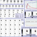
(a) At 2 months of life, a DMSA scan is performed after recurrent pyelonephritis. Scintigraphic imaging shows radiopharmaceutical uptake preserved in the left kidney and markedly reduced in the right one. Bilaterally, tracer distribution is not homogeneous due to multiple focal cortical defects with greater impairment of the right kidney (split renal function of right kidney was 29 %). Background signal is increased as for global insufficient renal function. At 18 months of life, a MAG3 renal scan confirms bilateral renal scars with preserved parenchymal function in the left kidney and severe hypofunction in the right one. Urinary excretion is mildly delayed in the left kidney, delayed and slow on the right, with evidence of radiotracer stasis which tended to resolve itself (b) and to further improve after orthostatism (gravity-assisted drainage test-1, d). Bilateral tracer accumulation is observed in the ureters, but both ureterograms show nonobstructive pattern. (e). Left renogram curve shows preserved slope of parenchymal phase and mildly slow elimination of tracer. Right renogram curve has a low width, showing reduced slope of parenchymal phase, delayed transit, and slow urinary excretion (c). The basic renogram shows a poor right split renal function, steady if compared to the previous DMSA scan. (f–g) IRC study is obtained only after several attempts, with a high filling bladder: performing an IRC study can sometimes be difficult due to poor child cooperation, but it is of crucial importance. In the IRC images, in fact, we can observe a regular and complete bladder emptying with bilateral VUR, more severe on the right side (f). Time-activity curves of kidneys reveal a bilateral radioactivity increment, synchronous with the bladder emptying, especially on the right (II–III scintigraphic grades on the left and III scintigraphic grade on the right) (g)
Case 6.6 Relapse of Monolateral Passive and Active Vesicoureteral Reflux Endoscopically Corrected, Secondary by Displacement of the Previously Injected Material
Female infant followed by pediatricians for suspected urinary tract infection. A cystography showed bilateral vesicoureteral reflux (Grade IV on the left, Grade I on the right), and a scintigraphic scan revealed a mild hypofunction of the left kidney. Thus, she underwent endoscopic treatment of vesicoureteral reflux, and baby was always asymptomatic with normal imaging follow-up. At 3 years of life, the child presented new infections of the urinary tract, and a radionuclide indirect cystography showed relapse of left passive and active vesicoureteral reflux (Fig. 6.6c, d). A new endoscopic treatment was performed, and at the cystoscopy, an evident displacement of the previously injected material was found. At the last clinical follow-up, the child remained asymptomatic.
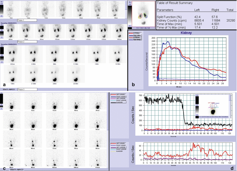
Fig. 6.6
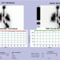
(a, b) By qualitative assessment of scintigraphic images, left kidney is smaller than normal with a slightly reduced renal uptake. Transit and urinary excretion of radiotracer from the kidney are slightly delayed and slow with evidence of mild pelvic stasis which tends to resolve itself during basal study. Right kidney shows an intense concentration of tracer in the renal parenchyma and a rapid washout (a). Left renogram curve (less wide than the contralateral one) shows preserved slope of parenchymal phase, transit slightly prolonged, and elimination of tracer from the kidney mildly slow. The shape of the right renogram curve is normal in all phases. The split renal function is slightly asymmetric (mildly reduced on the left) (b). (c, d) In order to obtain a diagnostic examination with improving sensitivity, IRC study was performed after oral hydration awaiting a better filling bladder and a further left pelvic stasis resolution. In the IRC images, we can observe persistence of left pelvic stasis, which slightly decreases and increases before bladder emptying. After a regular but incomplete bladder emptying (about 15 % of residual postvoiding activity), a high-grade VUR was detected on the left side (c). Time-activity curves of kidneys confirm a left radioactivity increment previously and synchronous with bladder emptying (passive and active VUR, II and III scintigraphic grades, respectively) (d). A good technical IRC performing is crucial to avoid false-negative findings or movement artifacts (especially when residual urine stasis is present)
Case 6.7 Indirect Radionuclide Cystography Evaluation After Endoscopic Vesicoureteral Reflux Treatment: Absence of Residual Reflux in Asymptomatic Child with Nephropathy
Female infant with febrile urinary tract infections. Renal ultrasonography previously performed showed no abnormalities while a cystography revealed bilateral vesicoureteral reflux (Grade III). A renal DMSA scintigraphy was performed showing multiple scarring in both kidneys, with greater impairment of the right one (split renal function of right kidney was mildly reduced). At 3 years of life, the child underwent endoscopic treatment of reflux. A control IRC was performed 1 year later, showing a steady hypofunction of right kidney and absence of residual reflux. Currently, she is asymptomatic.
Related posts:


Stay updated, free articles. Join our Telegram channel

Full access? Get Clinical Tree


