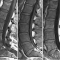
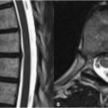
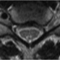
17 Neurodegenerative Disorders Age-related changes of the brain are commonly seen in clinical MRI. Findings include decreased SI in the basal ganglia from chronic iron deposition, atrophy, and foci of high SI on FLAIR and T2WI. These findings may be asymptomatic or pathologic. Diffuse atrophy is frequently visualized in Alzheimer disease as widening of the cortical sulci and dilatation of the ventricular system (ex vacuo hydrocephalus). The latter finding without corresponding sulcal widening, however, suggests a nonatrophic cause. In Alzheimer, atrophy of the hippocampal structures and temporal lobes may be especially prominent, as demonstrated in the sagittal T1WI and axial T2WI of Figs. 17.1A,B. Sulcal enlargement with diminution of the vermis and folia characterizes cerebellar atrophy as seen in Figs. 17.2A,B. Etiologies for cerebellar atrophy vary by age, with findings in a younger patient suggesting a primary cause like fragile-X syndrome, Friedreich, or spinocerebellar ataxia. Secondary atrophy may preferentially involve the vermis and pontocerebellar tracts and is seen in chronic phenytoin use (the etiology in Fig. 17.2) and alcoholism. Bilateral atrophy of the caudate nuclei (Fig. 17.3A, black arrows
![]()
Stay updated, free articles. Join our Telegram channel

Full access? Get Clinical Tree


