Discussion
The preferred terminology for describing disc “degeneration” and disc “displacement” has been the subject of debate and controversy for decades. Debate is still ongoing regarding the appropriate definition and usage of this terminology.
The most widely accepted recommended classification of lumbar disc pathology was formulated by a combined task force organized in the 1990s by the American Society of Spine Radiology (ASSR), the American Society of Neuroradiology (ASNR), and the North American Spine Society (NASS). The nomenclature recommendations formulated by this consensus task force were first published in 2001 and have been either endorsed, supported, or web-linked by the following organizations as of February 2003:
- 1
American Academy of Orthopedic Surgeons (AAOS)
- 2
American Academy of Physical Medicine and Rehabilitation (AAPM&R)
- 3
American Society of Neuroradiology (ASNR)
- 4
American Society of Spine Radiology (ASSR)
- 5
Joint Section on Disorders of the Spine and Peripheral Nerves of the American Association of Neurological Surgeons (AANS)
- 6
Congress of Neurological Surgeons (CNS)
- 7
European Society of Neuroradiology (ESNR)
- 8
North American Spine Society (NASS)
- 9
Physiatric Association of Spine, Sports, and Occupational Rehabilitation (PASSOR)
In spite of the broad support for usage of this nomenclature by the above organizations, confusion and controversy are still ongoing in regard to the proper usage of this terminology in the day-to-day clinical and imaging practice setting. Presented below is a summary of this standard system of nomenclature as used to describe lumbar disc displacement with additional comments regarding the limitations and controversy concerning this nomenclature system used for describing disc displacement. This nomenclature system was established based on the morphologic appearance of the lumbar intervertebral disc as viewed on images.
It is important to bear in mind that this terminology was specifically designed for describing lumbar disc pathology, although this terminology is often applied to cervical and thoracic disc disease as well. Some problems do arise when this system of nomenclature is applied to disc disease in the cervical and thoracic sections of the spine. Some author comments and observations are included below to address these issues and other controversies regarding this classification system where appropriate.
A summary of the existing disc classification with regard to disc displacement, along with our added commentary follows:
Disc Degeneration. The current classification of disc nomenclature includes disc degeneration, annular fissures (tears), and various types of disc herniations, with spondylosis deformans and intervertebral osteochondrosis being under the more general heading of degenerative or traumatic lesions of the intervertebral disc.
Comments: Controversy arises because there is no universal agreement as to when a normally aging disc becomes a pathologically deranged disc . Therefore no attempt is made in this section to discuss the nebulous concept of what constitutes disc degeneration as shown with imaging, from either a histologic or biomechanical perspective. This subject is dealt with in more detail in Chapter 17 concerning “internal disc derangement.”
Spondylosis deformans (vertebral osteophytosis) in general is believed to be the result of the normal aging process of the disc, although its exact etiology is still poorly understood. Anterior and lateral vertebral osteophytes are usually not considered pathologic. However, osteophytes, during their development, can become inflamed and when this occurs, can be a source of intense localized back pain. Posterior or posterolateral vertebral osteophytes are also often considered pathologic because they can encroach upon the spinal canal, spinal cord, or nerve roots within the spinal canal or neural foramina. The term intervertebral osteochondrosis would be more accurately phrased as discogenic vertebral osteochondrosis because the vertebral endplate irregularities, endplate sclerosis, and reactive vertebral marrow abnormalities in this condition are induced by the degenerative disc process. Annular tears are more appropriately called annular fissures because “tear” implies a traumatic etiology, which is probably not the cause of annular fissures in most cases.
Disc Bulging. A bulging disc is not a herniated disc by definition. A “bulging” disc, as defined in this classification system, is a disc that extends beyond the outer vertebral body margins by more than 3 mm circumferentially ( Figs. 5-1 and 5-2 ) . The modifier “generalized” or “circumferential” may be used if the disc margin extends greater than 180 degrees (greater than 50%) of the disc circumference. The term asymmetric disc bulge is used if the disc bulges greater than 180 degrees but less than 360 degrees of the disc circumference ( Fig. 5-3 ) .


Comments: If the disc extends beyond the outer vertebral margins by 360 degrees (100%) of the disc circumference, it should be called a bulging disc , which is the same as a diffuse disc bulge , a symmetric disc bulge , a generalized disc bulge , or a circumferential disc bulge . We prefer to use the term asymmetric disc bulge to describe a broad-based extension of the disc margin that involves greater than 90 degrees but less than 360 degrees of the disc circumference. However, the current classification defines an asymmetric disc bulge as one that involves greater than one half of the disc circumference and a broad-based disc protrusion as one that involves 90 to 180 degrees of the disc circumference. It is not possible with current imaging techniques to reliably differentiate a broad-based disc protrusion (which, by definition, histologically is “contained” by intact outer annular fibers) from a broad-based asymmetric bulging disc.
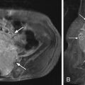
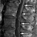
Disc Herniation. A disc herniation is a localized displacement of the disc margin beyond the usual confines of the disc. There are three major types of disc herniations based on the existing recommended classification, which is based on the morphologic imaging appearance of the disc as seen on computed tomography (CT) or magnetic resonance (MR): (1) disc protrusion, (2) disc extrusion, and (3) intravertebral disc herniation (Schmorl’s node), which causes vertebral endplate deformity ( Fig. 5-4 ) . A disc protrusion is defined as a localized contour abnormality involving less than 180 degrees (less than 50% of the disc circumference) with a wide base where the disc projects beyond the vertebral body margin in all imaging planes. To quote the original publication, disc “ protrusion is present if the greatest distance, in any plane, between the edges of the disc material beyond the disc space is less than the distance between the edges of the base, in the same plane. The base is defined as the cross-sectional area of disc material at the outer margin of the disc space of origin, where disc material displaced beyond the disc space is continuous with disc material within the disc space.” Disc protrusions that involve less than 90 degrees (less than 25%) of the disc circumference are considered to be “focal” or “localized” disc protrusions ( Figs. 5-5 to 5-7 ) . Disc protrusions involving 90 to 180 degrees of the disc circumference are referred to as broad-based disc protrusions using this standard nomenclature ( Fig. 5-8 ) .