Non-Hodgkin Lymphoma


Non-Hodgkin Lymphoma


Mary S. Huang

Nancy J. Tarbell

Howard J. Weinstein


Malignant lymphoma was described by Hodgkin in 1832 (1) and was distinguished from leukemia by Virchow in 1845 (2). Progress in treating children with non-Hodgkin lymphoma (NHL) mirrors the recognition of the systemic spread of the disease and underlying biology. Response to therapy and overall prognosis depend on the underlying histologic subtype, primary site, and the extent of disease (3,4). Historically, local therapy resulted in an overall survival of 10-30% (4). New multiagent protocols result in overall survivals of 70-95% (5, 6, 7, 8).

Childhood NHL is distinguished from adult NHL by differing frequencies of histopathologic types and by the greater frequency of extranodal presentations (9,10). The pediatric NHLs are mostly (more than 95%) high grade and include the following four major subtypes: B- and T-lymphoblastic lymphoma, Burkitt lymphoma, diffuse large B-cell lymphoma, and anaplastic large-cell lymphoma (ALCL) (Fig. 8.1). Many of these high-grade lymphomas disseminate noncontiguously, evolve into a leukemic phase, and involve the central nervous system (CNS) (10). The more common low-grade lymphomas seen in adults, such as follicular and marginal zone, are rare in children (9).


EPIDEMIOLOGY AND ETIOLOGY

The lymphomas are the third most common malignancy in children younger than 15 years of age. They are rare under the age of 3 years, peaking in incidence from age 7 to 11 years. There is approximately a 3:1 male:female ratio (3,4,6). Lymphomas account for approximately 10% of all childhood cancers: 60% are NHL and 40% are Hodgkin disease (11). There is geographic variation in the incidence of the NHLs. For example, in equatorial Africa, the Burkitt lymphoma accounts for almost 50% of all childhood cancers. In this setting, endemic Burkitt lymphoma is invariably positive for Epstein-Barr virus (EBV), in contrast to about 10% of cases of sporadic Burkitt lymphoma. However, both endemic and sporadic cases of Burkitt lymphoma have the same chromosomal translocations involving one of the loci encoding immunoglobin heavy or light chains and the c-myc oncogene. The exact role of EBV in the pathogenesis of Burkitt lymphoma and other malignancies is unknown. NHL occurs in association with congenital immunodeficiency syndromes such as X-linked lymphoproliferative syndrome, ataxia telangiectasia, Wiskott-Aldrich syndrome, and common variable immune deficiency disease, presumably caused by host defects in immunoregulation or gene rearrangement (12,13). Immunosuppressive therapy and acquired immunologic disorders including HIV also increase the risk of developing NHL (13). These are predominantly diffuse large B cell or Burkitt subtypes. Screening for HIV and other immunodeficiencies should be considered for all children with NHL, especially for those with B-cell lymphomas.


Figure 8.1 Denis Parsons Burkitt (1911-1993) was the son of an engineer. A Protestant raised in Northern Ireland, he lost an eye in a fight as a schoolboy. He graduated as a physician in 1935 and trained in surgery but had difficulty in obtaining surgical positions because of his visual loss. Eventually, he was posted as an Army physician in Africa and subsequently worked as a missionary physician. He characterized a previously undescribed aggressive head and neck cancer of childhood. Michael Epstein attended one of Burkitt’s lectures in England, suspected a viral origin, and requested a tissue sample. Epstein and Yvonne M. Barr isolated the virus (Epstein-Barr virus). Burkitt also described the crucial influence of dietary fiber on health and disease.


CLINICAL PRESENTATION

Several subtypes of NHL in children are associated with distinct clinical manifestations and sites of disease (
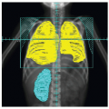
Table 8.1). Symptoms leading to diagnosis usually are of short duration. Approximately 25% of children with NHL have an anterior mediastinal mass (usually T lymphoblastic or diffuse large B cell) and present with wheezing, stridor, and cough
progressing to dyspnea. The majority of these patients are adolescents, and their presentation may pose a medical emergency (
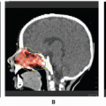
14). Patients with large anterior mediastinal masses are at risk of cardiac or respiratory arrest during general anesthesia or deep sedation. A careful workup including a chest computed tomography (CT) scan with airway measurements is essential (15) before attempting any procedures. The least invasive procedure (e.g., biopsy of a peripheral lymph node) should be carried out. If these procedures are not successful in providing a diagnosis, then a CT-guided needle biopsy of the mediastinal mass should be considered (16). In some clinical situations (e.g., orthopnea or significant airway narrowing), preoperative or preprocedure steroids should be considered for up to 48 hours. The use of steroids has largely replaced localized irradiation in this setting. Primary gastrointestinal involvement occurs in about 30% (usually Burkitt histology), commonly presenting as an abdominal mass with ascites, an “acute abdomen” from an intussusception, or rarely a malnutrition syndrome with colitis symptoms (6,17). The majority of children with Burkitt lymphoma presenting with an ileal-cecal intussusception have limited gastrointestinal involvement that is amenable to complete surgical resection (Murphy stage 2 or group A). In 20-30% of children, the head and neck, including the Waldeyer ring or cervical lymph nodes, is the site of origin. The remainder of patients have miscellaneous primary sites, including bone, breast, skin, epidural space, or noncervical lymph nodes (6). Involvement of the bone marrow at diagnosis occurs in 10-30% of patients with Burkitt and lymphoblastic lymphomas. Overt CNS involvement at diagnosis is not common but is mostly seen in children with advanced-stage Burkitt and lymphoblastic lymphomas. Children who develop Burkitt lymphoma in endemic areas of the world often have a mass in the head or neck region (especially jaw) in contrast to the abdominal presentation typical of nonendemic Burkitt lymphoma.


Table 8.1 Clinical and Biologic Features of NHL in Children


WHO Classification


Percentage of Cases


Common Primary Site


Immunophenotype


Chromosomal Changes


Genes Affected


Burkitt lymphoma


40


Abdomen or H/N


sIgM with light chain restriction CD10, CD19, CD20


t(8;14), t(2;8), or t(8;22)


Ig heavy- or lightchain genes and MYC


Diffuse large B-cell lymphoma (includes mediastinal)


20


Mediastinum, H/N, bone


sIgM > G (75%) CD79a, CD19, CD20, CD22


3q27, 8q24 and t(14;18) in adults


BCL-2 (adult, BCL-6)


B-lymphoblastic lymphoma


5


Bone, skin, H/N


TdT, CD10, CD19, CD79a


Hyperdiploid >50 (ALL)


TEL/AML1 (mostly in ALL)


T-lymphoblastic lymphoma


25


Mediastinum


TdT, CD2, CD3, CD4, CD5, CD7, and CD8, CD13 or CD33


14q11.2, 7q35, 7p14-15


TALI, TCR receptors, HOX 11, RBTN1


Anaplastic large-cell lymphoma


10


Skin, soft tissue, bone, mediastinum


CD30, EMA


t(2;5) or variant translocation


NPM-ALK


ALL, acute lymphoblastic leukemia; EMA, epithelial membrane antigen; H/N, head and neck; Ig, immunoglobulin; TCR, T-cell receptor; WHO, World Health Organization. Adapted from Jaffe ES, Harris NL, Stein H, et al. Classification of lymphoid neoplasms: the microscope as a tool for disease discovery. Blood 2008;112:4384-4399.


EVALUATION

A biopsy of a lymph node or mass or examination of pleural or ascitic fluid is necessary to establish the diagnosis. Although histology continues to be the primary determinant of therapy, morphology with immunohistochemistry is supplemented by cytogenetic, and molecular genetic studies. The workup includes complete blood cell count with differential, routine chemistries with electrolytes, calcium, phosphorus, magnesium, uric acid, liver and renal function tests, lactate dehydrogenase (LDH), chest radiograph, bone marrow aspirate and biopsy (18), and a lumbar puncture with cytocentrifugation. Abdominal, thoracic, and head and neck CT scans should be obtained and a magnetic resonance imaging (MRI) scan may provide additional information depending on the presenting site (19). Gallium and, more recently, positron emission tomography (PET) scans are now part of the initial staging for some patients with NHL and are particularly helpful in assessing response to induction therapy for diffuse large B-cell lymphoma and ALCL (20,21). Laparotomy is not indicated for staging and is performed only for abdominal presentations necessitating surgical intervention (e.g., ileal-cecal intussusception). There is no data to support surgical debulking of NHL in children.


STAGING AND CLASSIFICATION

Originally, the Ann Arbor staging system for the Hodgkin disease was used for NHL (see Table 7.1) (22). The usefulness of this system in pediatric NHL is limited, and an alternative staging system proposed by Murphy et al. (6) has become
widely accepted (Table 8.2). This system recognizes typical patterns of disease presentation and has greater prognostic utility than the Ann Arbor system. In Burkitt lymphoma and large B-cell lymphomas, another staging system developed by Patte et al. (23) is also used (Table 8.3). It classifies patients according to tumor burden and surgical resection.

The current most widely accepted histologic classification system is the World Health Organization (WHO) classification that was updated most recently in 2008 (24,25). This system has been validated as highly reproducible in international studies and has evolved to incorporate histology as well as immunohistochemistry, gene expression profiling, cytogenetic, molecular and clinical features.

As previously discussed, over 95% of the pediatric NHLs are high grade and fall into four major histologic subtypes (lymphoblastic lymphoma, Burkitt lymphoma, diffuse large B-cell lymphoma, and ALCL). T- and B-cell lymphoblastic lymphomas comprise about 30% of NHL in childhood; T cell is the more common type. Mature B-cell lymphoma includes Burkitt lymphoma (35-40% of cases) as well as diffuse large B-cell lymphoma (DLBCL) and primary mediastinal large B-cell lymphoma (PMBL). Together, the latter two comprise approximately 20% of NHL in childhood. In the current WHO classification, PMBL is considered distinct from other DLBCLs based upon its unique clinical, histologic, and molecular features. Other mature B-cell lymphomas, including pediatric marginal zone lymphoma, pediatric follicular lymphoma, and MALT lymphoma, are also recognized as distinct entities that rarely occur in childhood. Within the category of mature T-cell lymphoma, ALCL accounts for approximately 10% of NHL in childhood.

B-cell lymphoblastic lymphoma consists of cells with round or convoluted nuclei, fine chromatin, inconspicuous nuclei, and scant, faintly basophilic cytoplasm. In B-cell lymphoblastic lymphoma, the tumor cells are characteristically express TdT, CD10, CD19, and CD79a, which is identical to the most common immunophenotype of B-acute lymphoblastic leukemia (ALL) in childhood (24). The T-cell lymphoblastic lymphomas are also TdT positive, but express a combination of T-cell antigens (CD2, CD3, CD4, CD5, and CD8) similar to that of T-cell ALL. T-cell lymphoblastic lymphomas share many clinical and biologic features with T-cell ALL (26,27). When the bone marrow is involved with lymphoblasts (>25%), the distinction between lymphoma and leukemia becomes difficult. Subtle distinct biologic differences between the blasts in T-lymphoblastic leukemia and lymphoma are being recognized with newer molecular assays (28).


Table 8.2 Murphy and St. Jude Children’s Research Hospital Staging System for Childhood NHLa


Stage I


A single tumor (extranodal) or single anatomic area (nodal), with the exclusion of mediastinum or abdomen


Stage II


A single tumor (extranodal) with regional node involvement


Two or more nodal areas on the same side of the diaphragm


Two single (extranodal) tumors with or without regional node involvement on the same side of the diaphragm


A primary gastrointestinal tract tumor, usually in the ileocecal area, with or without involvement of associated mesenteric nodes onlyb


Stage III


Two single tumors (extranodal) on opposite sides of the diaphragm


Two or more nodal areas above and below the diaphragm


All the primary intrathoracic tumors (mediastinal, pleural, thymic)


All extensive primary intra-abdominal diseaseb


All paraspinal or epidural tumors, regardless of other tumor sites


Stage IV


Any of the preceding stages with initial CNS or bone marrow involvementc


aSee Refs. 6 and 10.

b A distinction is made between apparently localized gastrointestinal tract lymphoma and more extensive intra-abdominal disease. Stage II disease typically is limited to a segment of the gut with or without the associated mesenteric nodes only, and the primary tumor can be completely removed grossly by segmental excision. Stage III disease typically exhibits spread to para-aortic and retroperitoneal areas by implants and plaques in mesentery or peritoneum or by direct infiltration of structures adjacent to the primary tumor. Ascites may be present, and complete resection of all gross tumor is not possible.

c If marrow involvement is present initially, the number of abnormal cells must be 25% or less in an otherwise normal marrow aspirate with normal peripheral blood picture.


Table 8.3 Clinical Staging of B-Cell Lymphomas


Stage


Extent of Tumor


A


Resected stage I and abdominal stage II B Multiple extra-abdominal sites; nonresected stage I, II, III, and IV (CNS negative, BM > 25%)


C


Intra-abdominal tumor stage IV (CNS positive), BM > 25% (Burkitt leukemia)


From Patte P, Auperin A, Gerrard M, et al. Results of the randomized international


FAB/LMB96 trial for intermediate risk B-cell non-Hodgkin lymphoma in children and adolescents: It is possible to reduce treatment for the early responding patients. Blood. 2007;109:2773-2780.


CNS, central nervous system; BM, bone marrow.

Only gold members can continue reading. Log In or Register to continue

Stay updated, free articles. Join our Telegram channel

Jun 19, 2016 | Posted by in GENERAL RADIOLOGY | Comments Off on Non-Hodgkin Lymphoma

Full access? Get Clinical Tree

Get Clinical Tree app for offline access