Noninvasive Vascular Diagnosis


Noninvasive Vascular Diagnosis


Brian B. Ghoshhajra and Sanjeeva Prasad Kalva


Most vascular diseases can be diagnosed with a combination of clinical history and examination, relevant laboratory tests, and appropriate noninvasive tests. Such tests include the ankle-brachial index, segmental limb pressure and pulse volume recording measurements, Doppler assessment of blood flow, ultrasound evaluation of vessels, computed tomographic angiography (CTA), and magnetic resonance angiography (MRA). Computed tomography (CT) and magnetic resonance imaging (MRI) are now the first-line tests or replacements for diagnostic catheter angiography in many cases. The imaging test should be chosen based on the clinical problem. Other factors that may affect the choice of a particular test include cost, availability, urgency, pretest probability, and underlying contraindications. As with any radiologic tests, radiation exposure should be kept as low as reasonably achievable. In this chapter we briefly discuss the principles of ultrasonography, CTA, and MRA and their clinical applications.


Ultrasonography


Real-time grayscale ultrasonography is based on the principle of reflection of sound waves. The ultrasound transducer emits short pulses of high-frequency (1-10 MHz) ultrasound waves into the body and receives reflected echoes from the tissues. The computer analyzes the data and displays a grayscale image based on the intensity and timing of the reflected echoes. The reflection of sound waves depends on the differences in acoustic impedance of various tissues that form the interface. Large, smooth interfaces produce a high degree of reflection, which also depends on the orientation of the interface with respect to the angle of the incident ultrasound beam. In addition, small reflections, referred to as scatter, arise from multiple interfaces; these contribute to the diagnostic detail in the ultrasound images of solid organs. The scatter is not affected by the angle of the incident beam. Rayleigh scatter refers to diffuse scatter that occurs when the ultrasound beam interacts with tissues that are smaller than the wavelength of the ultrasound beam. This occurs commonly in the blood vessels, where red blood cells measure less than the wavelength of the ultrasound beam. Such scatter depends on many factors, including the number of scatterers (the red cells), size of the scatterers, and the ultrasound frequency. This scatter is responsible for Doppler evaluation of blood flow.


The Doppler effect refers to change in the perceived frequency when the source and/or the detector are moving. The red cells (the reflectors or the source) moving in the blood vessels change the frequency of reflected echoes (compared with the incident frequency of the ultrasound beam) depending on whether the motion is away or toward the transducer. Such change in the frequency is referred to as Doppler shift, which is governed by the following equation:


fD=fRfl=(2flVcosθ)/C


image

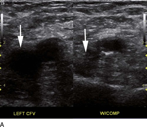
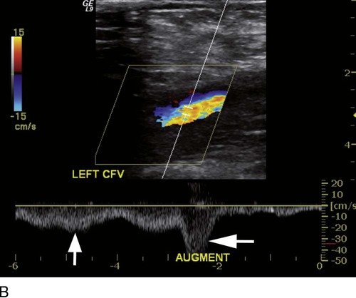
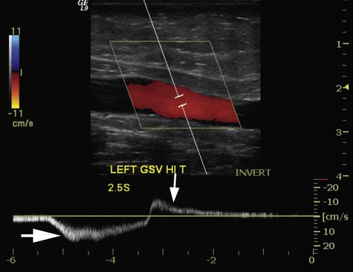
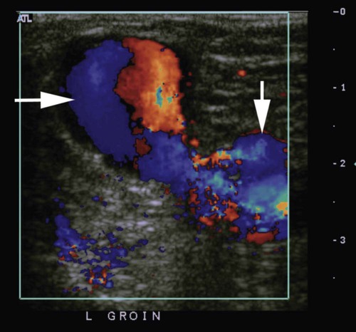
where fD = Doppler shift or Doppler frequency; fR = frequency of the reflected beam; fl is frequency of incident beam, V = velocity of the blood flow, θ = the angle between the direction of flow and the axis of the ultrasound beam, and C = velocity of the sound in tissues.


The computer detects the frequency of the reflected beam, and we know the frequency of incident beam and the velocity of sound in the tissues (1540 m/s). We can rewrite the equation to learn the velocity of the blood flow:


V=(fRfl)C/(2flcosθ)


image

When the angle of interrogation between the incident beam and direction of blood flow approaches 90 degrees, the cos θ approaches 0 and no Doppler shift is detected. In general, the angle of interrogation is kept between 30 and 60 degrees with the vessel lumen to receive reliable velocity measurements.


The Doppler shift is measured either with continuous or pulsed-wave Doppler. In continuous-wave Doppler, a transducer is placed over the area of interest and the reflected echoes are detected by another transducer. The ultrasound beam is emitted and received continuously. The magnitude of Doppler shift in the path of the beam is detected without spatial localization. Pulsed-wave Doppler imaging uses a single transducer that acts as both transmitter and receiver. Multiple short pulses of ultrasound beam are emitted, and the transducer turns to receiving mode. Generally, pulsed-wave Doppler is combined with grayscale ultrasound imaging (duplex ultrasonography), and thus the location of the ultrasound interrogation with the blood vessels can be identified and changed as required. With pulsed Doppler imaging, the maximum Doppler shift that can be measured depends on the pulse repetition frequency and the frequency of the transducer. When set conditions do not allow detection of the Doppler shift frequency, aliasing occurs, which is seen as wraparound Doppler spectrum on spectral wave form display or as complex colors on color flow imaging. Aliasing can be reduced by increasing the pulse repetition frequency or by using a low-frequency transducer.


The Doppler shift can be displayed in three different ways: color flow imaging, spectral waveforms, and audible sound. The mean velocity is color coded depending on whether the flow is toward or away from the transducer. The color flow maps are displayed over the grayscale image to provide better identification of the vessels. However, color flow imaging is less useful for quantification of blood flow and understanding the phasic nature of the blood flow. Spectral waveform display allows depiction of the direction of blood flow, phasic nature of blood flow, change in velocity over time (systolic, diastolic), and spectrum of velocities within the volume of interrogation. Audible sound is less useful in quantification and understanding the nature of the flow.


Power Doppler flow imaging differs from color Doppler imaging in that the intensity (or amplitude) of the Doppler shift is displayed in color, rather than the Doppler shift per se. It does not display the direction of blood flow. It is useful to detect “any flow” in the region of interest, and it is more sensitive in detecting blood flow and less dependent on the angle of interrogation.


Clinical Applications


Lower Extremities


Veins


Doppler evaluation of the venous system in lower extremities is usually performed to detect deep or superficial venous thrombosis (compression ultrasonography) or valvular insufficiency.1 Normal patent veins are anechoic and directly compressible with the transducer (Fig. e2-1, A). The Doppler spectrum demonstrates flow toward the heart, with respiratory variations in the flow pattern (the phasicity). Normal phasic flow in the distal veins suggests patent central veins. In addition, augmentation of flow can be demonstrated with distal compression (Fig. e2-1, B). An augmentation response usually suggests patent veins peripherally from the site of Doppler interrogation to the site of compression.


Acute deep venous thrombosis (DVT) is predisposed by hypercoagulable states (congenital or acquired), endothelial injury, and stasis of blood flow (Virchow triad). The common predisposing clinical conditions for DVT are malignancy, trauma, perioperative states, and postpartum, congenital, or acquired hypercoagulable states. Ultrasonography is highly sensitive in the diagnosis of acute DVT.2 Acute thrombosis results in an enlarged vein with intraluminal low-level echoes. The vein is noncompressible, and the Doppler spectrum shows no demonstrable flow. Subacute thrombus is seen as moderate high-level echoes in the lumen, with minimal or no enlargement of the vein. Chronic thrombosis is difficult to identify because the veins become small and may sometimes demonstrate channels of flow (partial recanalization of thrombus). Rarely, calcification of the vein may be seen as a focal highly echogenic area with distal acoustic shadowing.


Venous insufficiency predisposes to varicose veins, leg edema, venous stasis dermatitis, and venous ulcers. It can occur in the superficial or deep veins. Chronic venous insufficiency affecting the deep veins is usually predisposed by prior DVT. Venous insufficiency affecting the superficial veins may be related to prior thrombosis, but most often no cause is found. The elderly are predisposed to its development, and pregnancy and genetic factors also play a role. To plan appropriate therapy, it is necessary to document the site and degree of venous insufficiency and assess the anatomy of the superficial veins, perforator veins, and their relative contribution to varicosities. Venous insufficiency should ideally be assessed with the patient standing. A competent vein demonstrates flow toward the heart and transient reflux (<0.5 second) on a Valsalva maneuver or after distal compression. Reflux (Fig. e2-2) refers to reversal of flow direction (normal flow direction is toward the heart, from the superficial veins to the deep veins in case of perforator veins) and is graded depending on the length of time the flow reversal is observed.3 A longer duration of reflux implies more severe disease. Other parameters such as reflux velocity and the calculated reflux volume have been used to assess the severity of reflux.4


Arteries


Doppler evaluation of the arterial system is an adjunct to clinical evaluation and other noninvasive tests such as the ankle-brachial index, segmental limb pressures, and pulse volume recordings. Generally, duplex ultrasonography is requested to assess for focal stenoses or occlusions in the native arteries or bypass grafts.


Normally the arteries of the lower extremity demonstrate an anechoic lumen, with echogenic walls. Plaques are visualized as ill-defined low- or high-level echoes attached to the walls. Calcified plaques may demonstrate posterior acoustic shadowing. Doppler evaluation demonstrates a triphasic pattern of flow in all the arteries. Regional vasodilation due to exercise or inflammation in the leg leads to monophasic flow, with an increase in the diastolic component. However, this can be distinguished from poststenotic vasodilation because the latter demonstrates a tardus (slow upstroke) parvus (small amplitude) pattern. Although grayscale ultrasonography provides information regarding plaque burden, the hemodynamic significance is ascertained with Doppler imaging. An increase in peak systolic velocity of more than 100% compared with the proximal segment indicates a stenosis of at least 50% diameter. A higher degree of stenosis elevates the peak systolic velocity, and a fourfold increase in peak systolic velocity suggests severe stenosis. However, when the stenosis approaches 100%, the flow velocity may actually decrease. Occlusion can be seen as a lack of flow on color flow or Doppler imaging. Runoff vessels distal to an occlusion or significant stenosis demonstrate a tardus parvus pattern of flow on Doppler. This pattern of flow refers to slow upstroke of the systolic peak, with decreased peak systolic velocity. In addition, flow becomes monophasic, with increased diastolic flow due to regional vasodilatation. Acute vascular occlusions are usually secondary to emboli from the heart or due to vascular injuries. The emboli are seen as low-level echoes in the lumen, especially at the branch points, with a lack of flow demonstrated on color flow imaging. The proximal vessels may demonstrate a high-resistance flow pattern on the Doppler image—monophasic flow with no diastolic component.


Bypass grafts can be readily evaluated with ultrasound, owing to their superficial location. Ultrasonography is useful to assess patency of the graft, presence of thrombus, and occlusion. Occlusion is suspected when there is no Doppler signal detected in the graft. Evaluation for stenosis within the graft is imperfect because there are no standard criteria for grading severity; however, a twofold increase in peak systolic velocity compared to that of the proximal segment suggests a significant stenosis.


Ultrasonography may be requested to evaluate groin complications after endovascular interventions. These include hematoma, pseudoaneurysm, arteriovenous fistula, and arterial occlusion. Pseudoaneurysm is diagnosed when an anechoic mass with internal flow is identified adjacent to the puncture site. Generally a communication (i.e., “neck”) is identified between the mass and the artery (Fig. e2-3). If the communication is narrow, the pseudoaneurysm may be treated via ultrasound-guided thrombin injection. Arteriovenous fistula is diagnosed when the artery proximal to the fistula shows monophasic flow with increased diastolic flow, and the vein shows arterialized pulsatile flow. The actual site of communication is rarely seen by ultrasonography and may demonstrate elevated flow velocities. Arterial occlusions are secondary to thrombus formation or dissections. Ultrasonography may show intraluminal low-level echoes without flow on color flow imaging.


Upper Extremities


Veins


Ultrasound examination of the upper extremity veins may be requested as part of renal dialysis access management (dialysis fistula or graft planning, surveillance for venous stenosis, or for venous catheter placement), suspected DVT, or as part of a workup for recurrent pulmonary embolism. Respiratory variation in the flow may be less evident in the peripheral veins (brachial, ulnar, radial). DVT is diagnosed when the vein is enlarged, noncompressible, and demonstrates low-level luminal echoes without flow on color Doppler imaging. Paget-Schroetter syndrome refers to compression of the subclavian vein by the first rib or scalene muscles in the thoracic inlet; this condition predisposes to upper extremity venous thrombosis. The superficial veins are evaluated before a dialysis fistula is created to identify size, course, and patency. After fistula/graft creation, the central veins show arterialized monophasic flow. A stenosis is suspected when the peak systolic velocity increases by more than 100% compared with the proximal segment.


Arteries


An arterial study of the upper extremity may be required to evaluate suspected acute or chronic limb ischemia, assess patency of arteriovenous fistulas or grafts, and evaluate complications after arterial interventions. Acute arterial occlusions in the upper extremity are usually due to vascular injuries associated with trauma or intervention and emboli from the heart or proximal aneurysms (e.g., pseudoaneurysms of subclavian artery in association with thoracic outlet syndrome). These are seen as abrupt cessation of blood flow on color flow imaging, with low-level intraluminal echoes. Proximal vessels may demonstrate monophasic high-resistance flow on Doppler examination. Arterial stenosis is suspected if the peak systolic velocity increases by twofold or more compared with the proximal segment. Ultrasound may be requested to assess the location, size, and origin of pseudoaneurysms. Rarely, aneurysm of the distal ulnar artery may be identified in patients with hypothenar hammer syndrome. Dialysis fistulas and grafts are evaluated for patency, anastomotic stenosis, and venous outflow stenosis. Ultrasonography of the forearm arteries may be requested before harvesting the radial artery for coronary bypass. The radial and ulnar arteries are evaluated for patency and continuity of the palmar arches. If the palmar arch is intact, compression of one of the arteries should result in augmentation of flow, with increase in peak systolic velocity in the other artery.


Abdominal Vessels


Renal Artery


Renal artery stenosis is an uncommon but correctable cause of hypertension. The underlying etiology may be atherosclerosis (in the elderly), fibromuscular dysplasia (in the young), or may be congenital. Ultrasonography is used as a screening modality with variable success. Sensitivity varies from 0% to 98% among various studies.5,6 There are two approaches for the diagnosis of renal artery stenosis. The extrarenal arteries are evaluated for peak systolic velocity, and a velocity of more than 200 cm/s suggests a stenosis of more than 60% luminal diameter. In addition, the ratio of peak systolic velocity in the main renal artery and peak systolic velocity in the abdominal aorta (referred to as the renal/aortic ratio) is calculated. Renal artery stenosis is suspected if the ratio is more than 3.5. The second approach is to evaluate the intrarenal arteries by assessment of poststenotic flow dynamics. Renal artery stenosis is suspected if the intrarenal arteries demonstrate a tardus parvus pattern with an acceleration time (time to reach peak systolic velocity from baseline) more than 100 ms. Estimating the resistive index (RI) in the intrarenal arteries may be useful because an RI greater than 0.8 suggests a poor chance of recovery from renal dysfunction or hypertension after renal artery angioplasty. The main limitations of ultrasonography are difficulty in identifying the extrarenal arteries, inability to detect accessory renal arteries, and operator dependence. The extrarenal arteries may be better defined on ultrasonography with the use of contrast agents.


Transplant Kidney

Patients status post renal transplantation are evaluated for hydronephrosis, peritransplant fluid collections, and vascular pathology. Renal artery stenosis can occur at the anastomosis. Although there are no strict Doppler criteria to assess transplant renal artery stenosis, a peak systolic velocity of more than 200 cm/s in a straight segment of the artery usually suggests renal artery stenosis. Some recommend 300 cm/s as a cutoff point for diagnosing renal artery stenosis at the anastomosis. The iliac artery should also be evaluated to determine whether the increase in peak systolic velocity in the renal arteries is isolated or due to systemic hypertension. The intrarenal arteries may demonstrate poststenotic flow changes as described earlier. Intrarenal arteries are evaluated for resistive index; a value of 0.8 or more is suggestive of a parenchymal pathologic process such as rejection, acute tubular necrosis, or cyclosporine toxicity. Other vascular pathologic processes that may occur in the transplant kidney include renal vein thrombosis, arteriovenous fistulas, and pseudoaneurysms, which may be related to biopsy. Renal vein thrombus can be identified with Doppler imaging as lack of flow in the renal vein, with intraluminal low-level echoes. The main renal artery may demonstrate diastolic flow reversal in the presence of renal vein thrombus. Arteriovenous fistulas may be seen directly with color Doppler imaging, with an arterialized flow pattern in the draining vein. Pseudoaneurysm is seen as an anechoic mass with color flow within and is usually noted after a biopsy.


Mesenteric Arteries


Patients with chronic mesenteric ischemia present with postprandial pain and weight loss. There are excellent collateral pathways between the celiac axis, superior mesenteric artery, and inferior mesenteric artery. Unless two or more of these arteries are stenotic or occluded, it is rare to develop chronic mesenteric ischemia. A stenosis of more than 70% may be suspected if peak systolic velocities in the celiac axis and superior mesenteric artery exceed 200 cm/s and 275 cm/s, respectively.7 Indirect signs that suggest stenosis include identification of flow reversal in the vessels distal to the stenosis and enlargement of the inferior mesenteric artery. In addition, the normal expected postprandial increase in peak systolic velocity and diastolic flow may not be seen in the presence of severe stenosis of the superior mesenteric artery.8


Portal and Hepatic Veins


Evaluation of the portal venous system may be required for suspected portal hypertension, hepatic malignancies, and after decompressive surgery for portal hypertension and liver transplantation. Portal hypertension may result either from obstruction of flow or from excessive flow (hyperdynamic states). Portal venous obstruction is classically divided into three types: presinusoidal, sinusoidal, and postsinusoidal. In the presinusoidal type, the portal vein is obstructed before it communicates with the hepatic sinusoids, with the classic examples being portal vein thrombosis and compression of the extrahepatic portal vein by a mass or lymph nodes. Sinusoidal obstruction is usually from cirrhosis. Postsinusoidal obstruction occurs in hepatic veno-occlusive disease, pericardial constriction, and so on. Obstructive portal venous hypertension results in change in the direction of portal venous flow (flow directed away from the liver [hepatofugal]), development of portosystemic collaterals (e.g., splenorenal collaterals, recanalized umbilical vein, caput medusa, esophageal varices), and splenomegaly. Hyperdynamic portal hypertension is due to a fistula of the hepatic artery (or any mesenteric artery) to the portal vein or its tributaries. Intrahepatic arterioportal fistula results in hepatofugal flow in the portal vein with the development of portosystemic collateral vessels.


The normal portal vein measures 10 mm or less in diameter and shows an anechoic lumen. Doppler evaluation shows hepatopetal flow

Stay updated, free articles. Join our Telegram channel

Dec 23, 2015 | Posted by in INTERVENTIONAL RADIOLOGY | Comments Off on Noninvasive Vascular Diagnosis

Full access? Get Clinical Tree

Get Clinical Tree app for offline access