It is imperative for all imaging specialists to be familiar with detailed multiplanar CT anatomy of the paranasal sinuses and adjacent structures. This article reviews the radiologically relevant embryology of this complex region and discusses the region-specific CT anatomy of the paranasal sinuses and surrounding structures. Radiologists also need to know the clinical implications of identifying preoperatively the numerous anatomic variations encountered in this region and prepare a structured report according to the expectations of the referring clinician.
Anatomic concepts of the paranasal sinuses have been known since the late nineteenth and early twentieth centuries. These have assumed greater significance in recent times due to advances in functional endoscopic sinus surgery (FESS) and imaging technology. Multiplanar high-resolution CT (HRCT) of the paranasal sinuses provides a precise and reliable preoperative roadmap for the endoscopic sinus surgeon. All radiologists should be familiar with the 3-D anatomy of the paranasal sinuses and the anatomic variants that surgeons are likely to encounter. This article reviews the embryology of the paranasal sinuses and outlines the CT technique/protocols for imaging this region. CT anatomy of the nasal cavity and paranasal sinuses is described in detail together with the anatomic variants encountered in each region.
Knowledge of relevant embryologic events in paranasal sinus development can avoid pitfalls in diagnosis. Sinus pathologies in children younger than 4 years are uncommon except in the ethmoid sinuses because these are the only sinuses that are pneumatized at birth.
The mucosal lining over the nasal septum and the nasal turbinates is influenced by the nasal cycle, which is responsible for alternating changes in the turbinate sizes due to mucosal engorgement. This cyclic and physiologic enlargement of the turbinates alternates between both nasal cavities every 45 minutes to 1 hour and should not be mistaken for pathology.
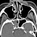
Septal pneumatization: pneumatization may occur anteriorly from the crista galli or posteriorly from the sphenoid sinus. Posterior septal pneumatization may occasionally narrow the sphenoethmoidal recess and impede access to the sphenoid ostium.
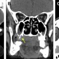
Pneumatized basal lamella: may be mistaken for an anterior ethmoidal air cell leading to incomplete exploration of the posterior ethmoid sinuses.
The suprabullar and retrobullar recesses can be identified while viewing the lamellar anatomy in the sagittal plane.
Rarely, the uncinate process may be entirely absent.
Bony margins of the infraorbital nerve canal may be dehiscent in up to 14% of cases, which exposes the nerve to sinus pathology.
Inadvertent intracranial penetration can be avoided during FESS by preoperative CT identification of a low ethmoid skull base, presence of a frontal bullar cell, Keros type 1 and type 3 olfactory fossae and asymmetric olfactory fossae.
E: Ethmoidal (anterior) artery anatomy
References
- 1. Ónodi A., and Thomson S.C.: The anatomy of the nasal cavity and its accessory sinuses: an atlas for practitioners and students. London: H.K. Lewis, 1895.
- 2. Gwaltney J.M., Phillips C.D., Miller R.D., et al: Computed tomographic study of the common cold. N Engl J Med 1994; 330: pp. 25-30
- 3. Figueroamon R.: Imaging anatomy in revision sinus surgery. In Kountakis S.E., Jacobs J., and Gosepath J. (eds): Revision sinus surgery. Berlin; Heidelberg (Germany): Springer, 2008. pp. 1-11
- 4. Aksoy E.A., Özden S.U., Karaarslan E., et al: Reliability of high-pitch ultra-low-dose paranasal sinus computed tomography for evaluating paranasal sinus anatomy and sinus disease. J Craniofac Surg 2014; 25: pp. 1801-1804
- 5. Dahmani-Causse M., Marx M., Deguine O., et al: Morphologic examination of the temporal bone by cone beam computed tomography: comparison with multislice helical computed tomography. Eur Ann Otorhinolaryngol Head Neck Dis 2011; 128: pp. 230-235
- 6. Bremke M., Leppek R., and Werner J.A.: Digital volume tomography in ENT medicine. HNO 2010; 58: pp. 823-832
- 7. Wormald P.J.: Imaging in endoscopic sinus surgery. In Wormald P.J. (eds): Endoscopic sinus surgery: anatomy, three-dimensional reconstruction and surgical technique, 3rd edition. New York: Thieme Medical Publishers, 2013. pp. 13-18
- 8. Lang J.: Clinical anatomy of the nose, nasal cavity and paranasal sinuses. New York: Thieme Medical Publishers, 1989.
- 9. Marquez S., Tessema B., Clement P.A., et al: Development of the ethmoid sinus and extramural migration: the anatomical basis of this paranasal sinus. Anat Rec (Hoboken) 2008; 291: pp. 1535-1553
- 10. Zeifer B.: Pediatric sinonasal imaging: normal anatomy and inflammatory disease. Neuroimaging Clin N Am 2000; 10: pp. 137-159
- 11. Som P.M.: Sinonasal cavity. In Som P.M., and Bergron R.T. (eds): Head and neck imaging, 2nd edition. St Louis: Mosby-Year Book, 1991. pp. 51-168
- 12. Sadler T.W.: Langman’s medical embryology. Philadelphia: Wolters Kluwer Health/Lippincott Williams & Wilkins, 2012.
- 13. Wolf G., Anderhuber W., and Kuhn F.: Development of the paranasal sinuses in children: implications for paranasal sinus surgery. Ann Otol Rhinol Laryngol 1993; 102: pp. 705-711
- 14. Anon J.B., Rontal M., Zinreich S.J., et al: Pre-and postnatal morphogenesis of the nose and paranasal sinuses. In Anon J.B., Rontal M., Zinreich S.J., (eds): Anatomy of the paranasal sinuses. New York: Thieme Medical Publishers, 1996. pp. 3-10
- 15. Som P.M., Shugar J.M.A., and Brandwein M.S.: Anatomy and physiology. In Som P.M., and Curtin H.D. (eds): Head and neck imaging, 4th edition. St Louis: Mosby, 2003. pp. 87-147
- 16. Kubal W.S.: Sinonasal anatomy. Neuroimaging Clin N Am 1998; 8: pp. 143-156
- 17. Beale T.J., Madani G., and Morley S.J.: Imaging of the paranasal sinuses and nasal cavity: normal anatomy and clinically relevant anatomical variants. Semin Ultrasound CT MR 2009; 30: pp. 2-16
- 18. Sarna A., Hayman A., Laine F., et al: Coronal imaging of the ostiomeatal unit: anatomy of 24 variants. J Comput Assist Tomogr 2002; 26: pp. 153-157
- 19. Wormald P.J.: Endoscopic sinus surgery: anatomy, three-dimensional reconstruction and surgical technique. New York: Thieme Medical Publishers, 2013.
- 20. Chong V., Fan Y., Lau D., et al: Functional endoscopic sinus surgery (FESS):what radiologists need to know. Clin Radiol 1998; 53: pp. 650-658
- 21. Zeinrich S.J., Mattox D.E., Kennedy D.W., et al: Concha bullosa: CT evaluation. J Comput Assist Tomogr 1988; 12: pp. 778-784
- 22. Cannon C.R.: Endoscopic management of concha bullosa. Otolaryngol Head Neck Surg 1994; 110: pp. 449-454
- 23. Stammberger H., Kopp W., and Dekornfeld T.J.: Special endoscopic anatomy. In Stammberger H., and Hawke M. (eds): Functional endoscopic sinus surgery: the Messerklinger technique. Philadelphia: B.C. Decker, 1991. pp. 61-90
- 24. Aribandi M., McCoy V.A., and Bazan C.: Imaging features of invasive and noninvasive fungal sinusitis: a review. Radiographics 2007; 27: pp. 1283-1296
- 25. Stammberger H.R., and Kennedy D.W.: Paranasal sinuses: anatomic terminology and nomenclature. Ann Otol Rhinol Laryngol Suppl 1995; 167: pp. 7-16
- 26. Lund V.J., Stammberger H., Fokkens W.J., et al: European position paper on the anatomical terminology of the internal nose and paranasal sinuses. Rhinol Suppl 2014; 24: pp. 1-34
- 27. Landsberg R., and Friedman M.: A computer assisted anatomical study of the nasofrontal region. Laryngoscope 2001; 111: pp. 2125-2130
- 28. Bolger W.E., Butzin C.A., and Parsons D.S.: Paranasal sinus bony anatomic variations and mucosal abnormalities: CT analysis for endoscopic sinus surgery. Laryngoscope 1991; 101: pp. 56-64
- 29. El-Shazly A.E., Poirrier A.L., Cabay J., et al: Anatomical variations of the lateral nasal wall: The secondary and accessory middle turbinates. Clin Anat 2012; 25: pp. 340-346
- 30. Bolger W.E., Woodruff W., and Parsons D.S.: CT demonstration of pneumatisation of the uncinate process. AJNR Am J Neuroradiol 1990; 11: pp. 552
- 31. Joe J.K., Ho S.Y., and Yanagisawa E.: Documentation of variations in sinonasal anatomy by intra-operative nasal endoscopy. Laryngoscope 2000; 110: pp. 229-235
- 32. Bolger W.E., Woodruff W.W., Morehead J., et al: Maxillary sinus hypoplasia: classification and description of associated uncinate process hypoplasia. Otolaryngol Head Neck Surg 1990; 103: pp. 759-765
- 33. May M., Sobol S.M., and Korzec K.: The location of the maxillary os and its importance to the endoscopic sinus surgeon. Laryngoscope 1990; 100: pp. 1037-1042
- 34. Vaid S., Vaid N., Rawat S., et al: An imaging checklist for pre-FESS CT: framing a surgically relevant report. Clin Radiol 2011; 66: pp. 459-470
- 35. Sathananthar S., Nagaonkar S., Paleri V., et al: Canine fossa puncture and clearance of the maxillary sinus for the severely disease maxillary sinus. Laryngoscope 2005; 115: pp. 1026-1029
- 36. Jog M., and McGarry G.W.: How frequent are accessory ostia? J Laryngol Otol 2003; 117: pp. 270-272
- 37. Wormald P.J.: Uncinectomy and middle meatal antrostomy including canine fossa puncture/trephine. In Wormald P.J. (eds): Endoscopic sinus surgery: anatomy, three-dimensional reconstruction and surgical technique, 3rd edition. New York: Thieme Medical Publishers, 2013. pp. 28-44
- 38. Earwacker J.: Anatomic variants in sinonasal CT. Radiographics 1993; 13: pp. 381-415
- 39. Wormald P.J.: Anatomy of the frontal recess and frontal sinus with three dimensional reconstruction. In Wormald P.J. (eds): Endoscopic sinus surgery: anatomy, three-dimensional reconstruction and surgical technique, 3rd edition. New York: Thieme Medical Publishers, 2013. pp. 45-80
- 40. Kew J., Rees G.L., Close D., et al: Multiplanar reconstructed computed tomography images improve depiction and understanding of the anatomy of the frontal sinus and recess. Am J Rhinol 2002; 16: pp. 119-123
- 41. Wormald P.J.: The agger nasi cell: the key to understanding the anatomy of the frontal recess. Otolaryngol Head Neck Surg 2003; 129: pp. 497-507
- 42. Kuhn F.A.: Chronic frontal sinusitis: the endoscopic frontal recess approach. Operative techniques. Otolaryngol Head Neck Surg 1996; 7: pp. 222-229
- 43. Stammberger H., and Lund V.: Anatomy of the nose and paranasal sinuses. In (eds): , 7th edition. London: HodderArnold, 2008. pp. 1315-1343
- 44. Meyers R.M., and Valvassori G.: Interpretation of anatomic variations of computed tomography scans of the sinuses: a surgeon’s perspective. Laryngoscope 1998; 108: pp. 422-425
- 45. Bhatti M.T., Schmalfuss I.M., and Mancuso A.A.: Orbital complications of functional endoscopic sinus surgery:MR and CT findings. Clin Radiol 2005; 60: pp. 894-904
- 46. Moon H.J., Kim H.U., Lee J.G., et al: Surgical anatomy of the anterior ethmoidal canal in ethmoid roof. Laryngoscope 2001; 111: pp. 900-904
- 47. Keros P.: On the practical value of differences in the level of the lamina cribrosa of the ethmoid. Z Laryngol Rhinol Otol 1962; 41: pp. 809-813
- 48. Ohnishi T.: Bony defects and dehiscences of the roof of the ethmoid bone. Rhinology 1981; 19: pp. 195-202
- 49. Kainz J., and Stammberger H.: The roof of the anterior ethmoid: a locus minoris resistentiae in the skull base. Laryngol Rhinol Otol 1988; 67: pp. 142-149
- 50. Som P.M., and Lawson W.: The frontal intersinus septal air cell: a new hypothesis of its origin. AJNR Am J Neuroradiol 2008; 29: pp. 1215-1217
- 51. Reddy U.M., and Dev B.: Pictorial essay: Anatomical variations of paranasal sinuses on multidetector computed tomography-How does it help FESS surgeons? Indian J Radiol Imaging 2012; 22: pp. 317-324
- 52. Stankiewicz J.A., and Chow J.M.: The low skull base: an invitation to disaster. Am J Rhinol 2004; 18: pp. 35-40
- 53. Rudmik L., and Smith T.L.: Evaluation of the ethmoid skull base height prior to endoscopic sinus surgery: a preoperative CT evaluation technique. Int Forum Allergy Rhinol 2012; 2: pp. 151-154
- 54. Weinberger D.G., Anand V.K., Al-Rawi M., et al: Surgical anatomy and variations of the Onodi cell. Am J Rhinol 1996; 10: pp. 1-6
- 55. Elwany S., Elsaeid I., and Thabet H.: Endoscopic anatomy of the sphenoid sinus. J Laryngol Otol 1999; 113: pp. 122-126
- 56. Wang J., Bidari S., Inoue K., et al: Extensions of the sphenoid sinus: a new classification. Neurosurgery 2010; 66: pp. 797-816
- 57. Maniscalo J.E., and Habal M.B.: Microanatomy of the optic canal. J Neurosurg 1978; 48: pp. 402-406
- 58. DeLano M.C., Fun F.Y., and Zinreich S.J.: Relationship of the optic nerve to the posterior paranasal sinuses: a CT anatomic study. Am J Neuroradiol 1996; 17: pp. 669-675
- 59. Osborn A.G.: The vidian artery: normal and pathologic anatomy. Radiology 1980; 136: pp. 373-378
- 60. Liu S.C., Wang H.W., Kao H.L., et al: Three-dimensional bone CT reconstruction anatomy of the vidian canal. Rhinology 2013; 51: pp. 306-314
Related posts:

Stay updated, free articles. Join our Telegram channel

Full access? Get Clinical Tree


