Fig. 20.1
Levels I–VI of the neck (and level VII, superior mediastinum)

Fig. 20.2
Level VI, anterior view
20.2 Level I: Submandibular
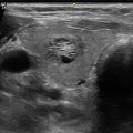
This group is very rarely involved in thyroid cancer. It consists of lymph nodes within the boundaries of the anterior and posterior bellies of the digastric muscles, the stylohyoid muscle, and the body of the mandible. Lymph nodes in this compartment are at greatest risk for harboring metastases from oral cavity, anterior nasal cavity, midface soft tissue, and submandibular gland malignancies. Inflammatory lymphadenopathy is also very common in level I, related to benign dental and oral cavity processes. Of note, the submandibular glands are located in level I, and these salivary glands are not infrequently affected by radioactive iodine therapy (Fig. 20.3).

Fig. 20.3
Level I lymph node (arrow) with adjacent submandibular gland
20.3 Level II: Upper Jugular
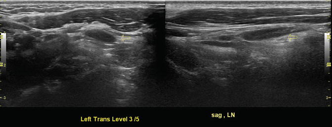
This group is comprised of lymph nodes located around the upper third of the internal jugular vein. Its boundaries are the skull base superiorly, inferior border of the hyoid bone inferiorly, lateral border of the sternohyoid and stylohyoid muscles anteromedially, and the posterior border of the sternocleidomastoid muscle posterolaterally. The spinal accessory nerve is within this compartment and subdivides this compartment into IIA which are nodes located anterior (medial) to the nerve and IIB which are nodes located posterior to the nerve. Level II nodes are at risk for harboring metastases from cancers arising from the oral cavity, nasal cavity, nasopharynx, oropharynx, thyroid, hypopharynx , larynx, and parotid gland (Fig. 20.4, Video 20.1).

Fig. 20.4
Benign level II lymph node (right transverse view)
20.4 Level III: Middle Jugular
This group consists of lymph nodes located around the middle third of the internal jugular vein. Its boundaries are the hyoid bone superiorly, cricoid cartilage inferiorly, lateral border of the sternohyoid muscle anteromedially, and posterior border of the sternocleidomastoid muscle posterolaterally. These nodes are at greatest risk for harboring metastases from cancers arising from the thyroid, oral cavity, nasopharynx, oropharynx, hypopharynx, and larynx (Fig. 20.5, Video 20.2).
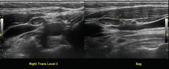
Fig. 20.5
Right level III benign lymph node , transverse and sagittal views
20.5 Level IV: Lower Jugular
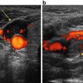
This group consists of lymph nodes located around the lower third of the internal jugular vein. Its boundaries are the cricoid cartilage superiorly, clavicle inferiorly, lateral border of the sternohyoid muscle anteromedially, and posterior border of the sternocleidomastoid muscle posteriorly. These nodes are at greatest risk for harboring metastases from cancers arising from the thyroid, hypopharynx, cervical esophagus, and larynx (Fig. 20.6).

Fig. 20.6
Right transverse level IV, somewhat amorphous but benign lymph node
20.6 Level V: Posterior Triangle
This group is comprised predominantly of the lymph nodes located along the lower half of the spinal accessory nerve and the transverse cervical artery, along with the supraclavicular nodes. The superior boundary is the apex formed by a convergence of the sternocleidomastoid and the trapezius muscles, the inferior boundary is the clavicle, the anterior boundary is the posterior border of the sternocleidomastoid muscle, and the posterior boundary is the anterior border of the trapezius muscle. Sublevel VA is separated from sublevel VB by a horizontal plane marking the inferior border of the arch of the cricoid cartilage. Sublevel VA includes the spinal accessory nodes, and sublevel VB includes the nodes following the transverse cervical vessels and the supraclavicular nodes. Level V nodes are at greatest risk for harboring metastases from cancers arising from the thyroid, nasopharynx, and oropharynx (Fig. 20.7).
 Ultrasound-Guided Laser Ablation
 Transcutaneous Laryngeal Ultrasonography: Clinical Variables
 Cytomorphology of Fine Needle Aspiration of Thyroid
 The Ultrasound Lexicon: Common Terminology for Thyroid and Parathyroid Sonography
 Ultrasound Characteristics and Ultrasound-Guided Fine-Needle Aspiration of Lymph Nodes in the Cervical Soft Tissues: A Radiology Perspective
 Pattern Recognition: Diffuse Thyroid Disease


Related posts:
Ultrasound-Guided Laser Ablation
Transcutaneous Laryngeal Ultrasonography: Clinical Variables
Cytomorphology of Fine Needle Aspiration of Thyroid
The Ultrasound Lexicon: Common Terminology for Thyroid and Parathyroid Sonography
Ultrasound Characteristics and Ultrasound-Guided Fine-Needle Aspiration of Lymph Nodes in the Cervical Soft Tissues: A Radiology Perspective
Pattern Recognition: Diffuse Thyroid Disease
Stay updated, free articles. Join our Telegram channel
Full access? Get Clinical Tree
