Fig. 44.1
Tubercle of Zuckerkandl: surgical specimen with arrows pointing to bilateral tubercles
44.5 White Knight
A hyperechoic (“white”) nodule in Hashimoto’s thyroiditis thought to represent regenerative benign tissue nodule appearing within the inflammation of the thyroid. This entity is discussed also in Chap. 16 (Lupo) and described in detail in the following two publications. Please see Video 44.4 [5, 6].
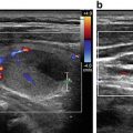
44.6 Starry Starry Night
A dense collection of microcalcifications that appear to “sparkle” like stars in the nighttime sky. This is a sonographic sign that is highly suspicious for thyroid cancer. The microcalcifications represent psammoma bodies, which in turn are involuted and necrotic papillae of papillary thyroid cancer. Please see Video 44.5 [7].
44.7 Comet Tail Artifact
A reverberation of sound waves caused by inspissated colloid and a marker of a benign process. This sonographic sign is often misinterpreted as microcalcifications. A key feature to recognize is that colloid fragments precipitate in cystic structures. In contrast, microcalcifications associated with thyroid cancer occur in solid nodules. Please see Videos 44.6 and 44.7 [8–10].
44.8 Giraffe Pattern
A sonographic pattern seen with Hashimoto’s thyroiditis and described by Bonavita and colleagues as a “giraffe” pattern similar to the two-tone coloring of a giraffe (see cartoon example). The ultrasound appearance is described as globular, block-like, or plate-like areas of hyperechogenicity surrounded by linear thin areas of hypoechogenicity [5] (Fig. 44.2a–d).
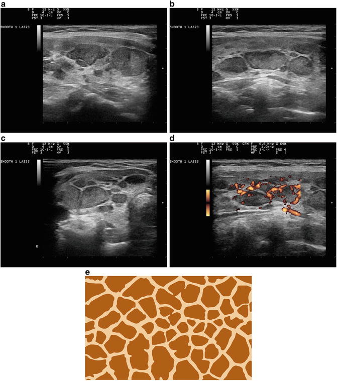
Fig. 44.2
(a–d). Giraffe pattern. Images are courtesy of the personal image collection of Dr. John S. Abele, Sacramento, California. (e) Giraffe skin pattern
44.9 Three Circle Sign
This lexicon terminology was first reported in the textbook, Diseases of the Parathyroid Glands , published by Springer in 2012. It describes the arrangement of circular cross-sections of the internal jugular vein (IJ), common carotid artery (CA), and the enlarged parathyroid gland when viewed by ultrasound in a transverse orientation. Figure 44.3 illustrates this orientation. The unique aspects of the “three circle sign” include the following: the uniform hypoechoic echogenicity of all three structures (a thyroid nodule may not exhibit this) and the absence of a rim of normal thyroid tissue between the endocrine gland and the carotid artery (a thyroid nodule will typically be within the gland parenchyma and the abnormal parathyroid gland is almost always extracapsular to the thyroid). The arrangement can be observed with both superior and inferior enlarged parathyroid glands. Video 44.8 demonstrates in color Doppler mode a very small caliber IJ at the left of the screen, the CA in the center of the video and the hypoechoic enlarged parathyroid gland at the right of the screen. Video 44.9 illustrates all three round structures, with a valve seen fluttering within the IJ at the left of the screen [11].

Fig. 44.3
Three circle sign
44.10 Pseudonodules and Cobblestones
The presence of thyroiditis can distort the thyroid architecture into globular arrangements that may appear to be discreet nodules but are really a conglomerate of round shapes of varying sizes that reflect inflammation and compartmentalization of tissue. These round areas, especially if they represent the pattern of the entire thyroid gland, should not be viewed as discreet nodules and should not undergo biopsy. Please see Figs. 44.4 and 44.5, and Videos 44.10, 44.11, 44.12, and 44.13 [12, 13]