Fig. 27.1
Typical appearance of a parathyroid adenoma seen as a (a) solid oval lesion with well-defined margins and hypoechoic relative to the thyroid gland (calipers in image). Color Doppler shows the typical arborization of blood supply in the parathyroid adenoma (b)

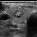
Fig. 27.2
Enlarged extra-thyroidal artery (arrow) seen arising from the inferior thyroidal artery and supplying the parathyroid adenoma (*)
27.2 Elusive Adenoma
While usually scanning for parathyroid adenomas is straightforward, identification of adenomas may be challenging in some cases. Some of the atypical appearances and abnormal locations are described below along with the imaging pitfalls and scanning technique to be used.
27.3 Atypical Findings
Although most adenomas are less than 2 cm in size, large adenomas greater than 5 cm can occur [4]. Most adenomas are oval in shape; however, some can have an asymmetric shape as well (Fig. 27.3) especially when large. Occasionally, parathyroid adenomas may be echogenic or heterogeneous in echotexture rather than displaying the usual homogenous hypoechoic appearance. This is especially possible when the adenomas are larger than 2 cm (Fig. 27.4, Video 27.4) [5]. Cystic adenomas are seen to occur in 2 % of cases and may be difficult to differentiate from rare true parathyroid cyst (Fig. 27.5, Video 27.5) [4]. In patients with parathyroid hyperplasia, all four glands are enlarged to varying degrees with the glands being usually smaller in size than in patients with parathyroid adenomas (Fig. 27.6). Hence, they are not as readily visible [6]. Hyperplasia is more commonly seen in patients with familial syndromes such as Multiple Endocrine Neoplasia (MEN) 1 and MEN 2A [5].


Fig. 27.3
Large parathyroid adenoma measuring 4.5 cm seen posterior to the thyroid with a homogenous hypoechoic appearance on Bmode image (a) and peripheral vascularity on Color Doppler (b)
Fig. 27.4
Large parathyroid adenoma with a heterogenous appearance and lobulated margins. Note the cystic areas (arrow) in the adenoma (a) and prominent vascularity on color Doppler (b)
Fig. 27.5
Superior parathyroid adenoma with a partially cystic appearance (calipers in image)
Fig. 27.6
Parathyroid hyperplasia in primary hyperparathyroidism. 54 year old female with a persistently elevated parathyroid hormone level. Ultrasound showed small right superior (calipers in a), left superior (arrow in b) and left inferior (arrow in c) hypoechoic nodules posterior to the thyroid gland suggesting multiple adenomas or parathyroid hyperplasia
Multiple adenomas are seen to occur in 5 % of patients (Fig. 27.7, Video 27.6). In these cases, the surgical management will change from unilateral minimally invasive operation to bilateral neck dissection; identification of these patients is crucial. Hence, it is imperative that all the four quadrants and potential ectopic locations are evaluated, even if one abnormal parathyroid gland has already been identified [7–9].

Fig. 27.7
Multiple parathyroid adenomas . Two large hypoechoic nodules seen posterior to the right thyroid gland in a patient with elevated parathyroid hormone levels (arrow and double arrows) suggesting multiple adenomas
Intrathyroidal adenomas are impossible to differentiate from thyroid nodules, however should be suspected in patients with non-localized parathyroid adenomas and presence of primary hyperparathyroidism. Intrathyroidal adenomas are seen in only 6 % of cases [10]. On imaging, a homogenously hypoechoic nodule is seen within the thyroid capsule (Fig. 27.8). A large feeding artery may be seen; however, in the review by Yabuta et al., feeding artery was not commonly seen and only an echogenic line between the parathyroid adenoma and the surrounding thyroid tissue on the ventral surface of the parathyroid gland was seen [11].

Fig. 27.8
Intrathyroidal parathyroid adenoma . Hypoechoic nodule seen along the posterior aspect of the left thyroid lobe on B-mode image (a). Color Doppler image did not show any polar vessel blood supply (b). Presence of echogenic line in between the adenoma and the thyroid (arrow) suggested a parathyroid adenoma . This was confirmed by fine needle aspiration
27.4 Abnormal Location
The parathyroid glands develop from the third and fourth pharyngeal pouches and usually two glands develop on each side, one superior and one inferior. This can range from 2 to 6 glands with some authors reporting up to 12 glands [12, 13]. Since the thymus and the inferior parathyroid glands arise from the third pharyngeal pouch and since the thymus descends into the mediastinum, ectopic inferior parathyroid glands may be located anywhere along this pathway (Fig. 27.9, Videos 27.7 and 27.8). The superior parathyroid glands arise from the fourth pharyngeal pouch along with part of the thyroid gland [14].

 Ultrasound-Guided Laser Ablation
 Transcutaneous Laryngeal Ultrasonography: Clinical Variables
 Cytomorphology of Fine Needle Aspiration of Thyroid
 The Ultrasound Lexicon: Common Terminology for Thyroid and Parathyroid Sonography
 Feature Illustration: Vascularity
 Pattern Recognition: Diffuse Thyroid Disease


Related posts:
Ultrasound-Guided Laser Ablation
Transcutaneous Laryngeal Ultrasonography: Clinical Variables
Cytomorphology of Fine Needle Aspiration of Thyroid
The Ultrasound Lexicon: Common Terminology for Thyroid and Parathyroid Sonography
Feature Illustration: Vascularity
Pattern Recognition: Diffuse Thyroid Disease
Stay updated, free articles. Join our Telegram channel
Full access? Get Clinical Tree
