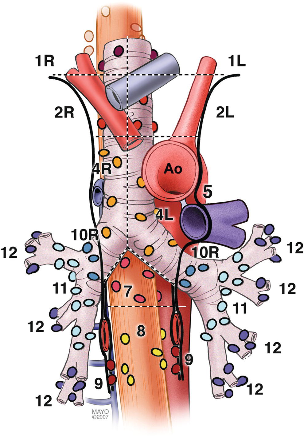
Juan Corral1, Sebastian Fernandez‐Bussy2, and Michael B. Wallace1 1 Division of Gastroenterology and Hepatology, Mayo Clinic College of Medicine, Jacksonville, FL, USA 2 Division of Pulmonary Medicine and Critical Care, Mayo Clinic College of Medicine, Jacksonville, FL, USA The mediastinum is a common anatomical location for lymph node (LN) metastases in lung cancer as well as many other malignant and inflammatory conditions. The presence and specific location of mediastinal LN metastases in non‐small cell lung cancer (NSCLC) dictates therapy with surgery for localized disease, combination therapy when contralateral LNs are involved, and palliative therapy when contralateral LNs and metastases are encountered. Unfortunately, cross‐sectional imaging with computed tomography (CT), magnetic resonance imaging (MRI), or positron emission tomography (PET) alone is not adequate to confirm a diagnosis; thus, a tissue sample is preferred. Recently, it has been suggested that the use of endoscopic ultrasound‐guided fine needle aspiration (EUS‐FNA) associated with endobronchial ultrasound‐guided transbronchial fine needle aspiration (EBUS‐TBNA) can adequately sample LNs in the mediastinum, avoiding the need for a futile surgery. The purpose of this chapter is to provide the basic anatomical information as well as technical maneuvers used to investigate the mediastinum successfully. The LNs in the mediastinum were classified in different stations based on surgical and anatomical landmarks for the purpose of staging lung cancer but this schema is now widely used in other chest diseases (Figure 3.1). The LNs with their respective stations and corresponding anatomical locations are described in Table 3.1. EUS‐FNA is usually best suited to sample LNs adjacent to the esophagus which runs posterior to the trachea. Because of ultrasound artifacts created by the air‐filled trachea, lesions immediately anterior to the trachea are not well seen. EUS‐accessible stations include 2L, 2R, 4L, 4R, 5, 7, 8, 9, and, sometimes depending on the size, station 6. On the other hand, EBUS‐TBNA can target LNs either anterior or lateral to the trachea to the level of the carina, and alongside the left and right bronchial tree including stations 2L, 2R, 4L, 4R, 7, 10, and 11. Although both procedures overlap in stations 2 L/R, 4 L/R, and 7, in other stations they are complementary, and in combination allow nearly complete mediastinal access. Radial and curvilinear array echoendoscopes are available (Figure 3.2), with scanning radius ranging from 270–360 degrees for radial to 100–180 degrees for the linear echoendoscope. These scopes have standard accessory channels (2.0–2.8 mm) and larger accessory channels (3.7 mm) capable of delivering needles and other therapeutic devices such as a 10 French (Fr) plastic stent. EUS can use several types of needles: 19 gauge (G), 22 G, and 25 G for FNA, as well as Tru‐cut needles for core biopsy. The needle is occluded with a stylet during passage through the gastrointestinal tract wall and bronchial wall to minimize contamination from passage through those structures. EBUS equipment comprises a curvilinear array echoendoscope with an outer diameter of 6.7 mm and a biopsy channel of 2 mm. The ultrasonic frequency is 7.5 MHz with a penetration depth of 4–5 cm, making it well suited for FNA of LNs and lung masses through the trachea and bronchi. A 22 G needle is used to perform TBNA in the same manner as in EUS. Both systems have integrated oblique‐viewing optics to guide intubation and limited inspection. Figure 3.1 Mediastinal lymph node stations. The initial examination can be performed with either the radial or linear array echoendoscope; however, the linear scope is required to perform FNA. Given the obvious efficiencies, we prefer to use a single linear echoendoscope for both imaging and FNA. The balloon should be deflated or inflated only slightly to provide good acoustic coupling with the tissue. The mediastinum is imaged by first finding the descending aorta starting at the cardia. The examination can be performed by rotating 360 degrees from the cardia, then withdrawing the shaft 4–5 cm and performing another rotation. Alternatively, one can survey from the cardia to the cervix, then rotating 90 degrees and repeating the maneuver until the whole mediastinum is examined. It is useful to use the following five stations as described by Deprez (Videos 3.1.1–3.1.3). For radial examination, see Video 3.2. Table 3.1 Mediastinal lymph node stations with their anatomical correlations. Figure 3.2 Types of echoendoscopes: (a) linear probe; (b) endobronchial probe; (c) radial probe. The descending aorta is a large echo‐poor longitudinal structure on linear array with a bright deep wall due to the air interface with the left lung. Clockwise rotation will sequentially image left lung, left pleura, left atrium, right lung, right pleura, azygos vein, and spine. The azygos vein can be localized by rotating approximately 30 degrees counterclockwise from the descending aorta. It is a thin echo‐poor structure that can be followed proximally to its union with the superior vena cava. This is the area of LN stations 8 and 9 (Figure 3.3). Figure 3.3 Lymph node at station 8 (between calipers). Figure 3.4 Subcarinal station (station 7). At approximately 30 cm the subcarinal area is easily recognized by finding the left atrium, a large hypoechoic structure with cardiac motion, and pulling back until it disappears on the left edge of the screen. Then one should have the pulmonary artery in the right portion of the screen (Figure 3.4). Slight movements to the right and left have to be performed to completely interrogate this station. This is the area of LN station 7 (Videos 3.3 and 3.4). The azygos arch is located at 24–25 cm from the incisors. The aortopulmonary (AP) window (station 5) is situated between the aortic arch and the pulmonary artery. The AP window is found by following the aorta cephalad until its arch, rotating clockwise approximately 90 degrees, then advancing 1–2 cm with slight tip up of the echoendoscope. The aorta will be the echo‐poor structure on the right and the pulmonary artery will be to the left; the AP window is the space between the two just outside the AP ligament (which is not seen by EUS). The 4L region is immediately medial (close to the esophagus and EUS scope) to the AP window (Figure 3.5). Alternatively, from the subcarinal area, rotating 90 degrees counterclockwise, crossing the left main bronchus and pulling it back 2–3 cm will put you in the same location. Further withdrawal of the echoendoscope with slight rotation will show the origin of the left subclavian artery. Occasionally, the left carotid artery can be seen above the brachiocephalic (innominate) vein. Figure 3.5 Aortopulmonary window station (stations 4L, 5 and 6). AO, aorta; L node; lymph node; PA, pulmonary artery. Between 22 and 24 cm from the incisors, the superior part of the lungs can be imaged along with the trachea, cricoid bone, jugular veins, and carotid arteries. At 17–20 cm from the incisors, the inferior thyroid gland can be imaged as well as cervical LNs, which can be targeted for FNA. For radial scanning, one should enter the stomach, inflate the balloon, and pull back the scope until the GE junction. The aorta with be visualized as a large round echo‐poor structure. The aorta should be rotated to the 5 o’clock position, and the spine will be at 7 o’clock with the azygos vein in the middle. Then the balloon should be deflated and a slow pull‐back is performed observing all the mediastinal areas. It is important to note that, for a complete lung cancer staging with both radial and linear, the celiac axis, the left adrenal, and the left lobe of the liver should be surveyed. This is easily accomplished with a slight tip down on the echoendoscope and and then pushing it into the stomach; the celiac take‐off can be seen and the left adrenal can be seen at the 6 to 4 o’clock position with its usual “seagull” appearance. For the liver, one goes to the gastric antrum, aspirates all the air, inflating the balloon, and then tip up with a slow pull‐back. Just like EUS, EBUS can be performed with conscious sedation, deep sedation, or general anesthesia. The tip of the scope is placed under direct visual contact in different positions, starting from the segmental bronchi to the trachea usually scanning 90–120 degrees every 0.5–1 cm. The diameter of the EBUS scope (6–7 mm) precludes passage beyond the second or third airway generation. EBUS‐TBNA is performed under the same principles as EUS‐FNA. As mentioned before, this technique is particularly useful for LN stations 2, 4, 7, 10, and 11 (Figure 3.6). The upper and middle third of the esophagus can be visualized from the upper and lower trachea respectively Currently, we have different sizes of needle available, including 19, 21, 22, and 25 G. It is important to note that all right tracheal lymph node stations are found from the left lateral border of the trachea to the right aspect of the trachea. Figure 3.6 Endobronchial ultrasound (EBUS) images of common mediastinal stations (4L, 7 and 11L). The esophagus and vertebral bodies can be visualized posteriorly from the upper and lower trachea. EUS‐FNA and EBUS‐TBNA are highly safe procedures in experienced hands, with a complication rate of 0.8%. A major safety precaution with FNA is to visualize the entire length of the needle and to use color Doppler to avoid any blood vessels in the needle path. EUS‐FNA and EBUS‐TBNA are complementary procedures with a high degree of sensitivity and specificity for diagnosing and staging benign and malignant diseases of the chest. Careful attention to technique must be applied to prevent the omission of important clinical information.
3
Normal Mediastinal Anatomy by EUS and EBUS
Introduction
Anatomical definitions
Equipment
Endoscopic ultrasound technique
Linear scanning
Level
Anatomical correlation
Superior mediastinal lymph nodes
1
Highest mediastinal
2
Upper paratracheal
3
Prevascular and retrotracheal
4
Lower paratracheal (including azygos nodes)
Aortic lymph nodes
5
Aortopulmonary (AP) window or subaortic
6
Para‐aortic (ascending aorta and phrenic)
Inferior mediastinal lymph nodes
7
Subcarinal
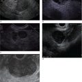
8
Paraesophageal (below carina)
9
Pulmonary ligament
N1 lymph nodes
10
Hilar
11
Interlobar
12
Lobar
13
Segmental
14
Subsegmental 
Inferior posterior mediastinum


Subcarinal area
Aortic arch area

Cervical area
Thyroid gland
Radial scanning
Endobronchial ultrasound
EBUS anatomical landmarks

Complications and safety
Conclusions
Related posts:


Stay updated, free articles. Join our Telegram channel

Full access? Get Clinical Tree


