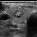
Fig. 9.1
Transverse anatomy of the neck through the thyroid
Knowing the relevant anatomic structures and their relationships to each other will make learning and performing thyroid and parathyroid ultrasound more straightforward and enjoyable. Being able to expertly and confidently conduct and interpret the ultrasound exam will also set your patients’ minds at ease.
Exams should follow a standard and sequential approach. Proper patient positioning will lift the thyroid up above the clavicles and allow the ultrasonographer to obtain a more complete exam. This is achieved by placing a shoulder roll under the patient in the supine position, which achieves moderate neck extension. The best place to begin the exam is in the midline directly over the trachea, as it is the easiest landmark to identify by physical exam. A high-frequency probe is preferred for head and neck ultrasound, as it provides excellent image resolution of the structures of interest, which are usually no more than 5–6 cm deep. When saving images, the laterality and orientation of each image should be clearly labeled.
The trachea has a characteristic appearance caused by the echogenic cartilage rings that surround the anterior and lateral aspects of this otherwise hollow tube (Fig. 9.2, Video 9.1). Placing the probe transversely directly over the trachea, one will quickly see the very bright hyperechoic stripes corresponding to the tracheal cartilage rings. Moving the probe in a caudal direction from the thyroid cartilage, one first encounters the cricoid cartilage, then followed by a number of cartilage rings. The patient’s body habitus and neck length will determine how many rings can be seen, but it is usually not more than a couple before the manubrium is encountered. The lumen of the trachea itself appears nearly black, as it is a hollow structure, which does not reflect any of the ultrasound waves passing through it. There are internal echoes present in the lumen that occur as an artifact related to the cartilage rings. The most superficial structures encountered are the skin and subcutaneous tissue (Fig. 9.3). The dermis is the most superficial layer. It is 1–2 mm in thickness and has a similar echogenicity to the thyroid gland. Just deep to it is a layer of subcutaneous fat, its width variable according to the patient’s body habitus. The echogenicity of this fatty layer is brighter than the dermis and is starkly contrasted with the muscle layers that lie beneath it. To either side of the midline, just below the subcutaneous fat, lie the strap muscles, specifically the sternohyoid more superficially and medial and the sternothyroid , deeper and more lateral. In the transverse view, each of them is the shape of a convex lens. They are quite hypoechoic relative to the overlying fat and underlying thyroid, and each is enveloped by a fine white line. The deeper sternothyroid extends from the sternum up to the thyroid cartilage, and the more superficial sternohyoid is essentially seen the entire length of the neck, as it inserts upon the hyoid bone.

Fig. 9.2
Trachea in cross section
Fig. 9.3
Axial image of superficial structures. S = skin, PM = subcutaneous fat, SH = sternohyoid muscle , ST = sternothyroid muscle , SCM = sternocleidomastoid muscle, I = isthmus of the thyroid, LL = left lobe of the thyroid, CA = common carotid artery , JV = internal jugular vein
Immediately posterior to the strap muscles sits the thyroid gland (Fig. 9.4). Its two lobes straddle the trachea, connected by the narrow isthmus. The normal thyroid gland is homogeneous in echotexture. It appears hyperechoic as compared to the overlying strap muscles. Each lobe sits snuggly between the trachea medially and the carotid artery laterally. The two lobes normally appear as mirror images of each other and usually are quite similar in size. Because the esophagus normally resides slightly to the left of the midline, it is frequently seen as an additional structure posterior to the left thyroid lobe (Fig. 9.5).


Fig. 9.4
Axial view through the thyroid and adjacent structures
Fig. 9.5
Structures found posterior to the thyroid
The contours of the thyroid are usually very smooth and quite discrete. Each lobe is bordered by the trachea medially, strap muscles anteriorly, and the carotid artery laterally. Posteriorly, there is the fat and lymphatic tissue that fills the central compartment between the spine and the thyroid. Because that tissue does not lie within any defined structure, the posterior border of the thyroid is often less well defined. The fatty tissue behind the thyroid usually appears heterogeneous and can be darker or brighter than the overlying thyroid parenchyma.
Thyroid size varies from patient to patient, but in general, in adults, the width (W) or transverse diameter and depth (D) or anterior-posterior diameter are quite similar, generally between 1.5 and 2.5 cm. The height or length (L) of the thyroid lobe is typically 4–5 cm. The thickness (anterior-posterior diameter) of the isthmus is usually less than 5 mm. In most patients the normal lobes are quite similar in size; however, rarely there will be a rather undeveloped lobe unilaterally. The measurement of the thyroid lobes and focal lesions should be performed in a standard fashion, and the order should be specified, so that comparisons of nodules and sizes of lobes can be made. For example, these authors report the dimensions as W × D × L, whereas many ultrasound labs will report dimensions as D × W × L. Many sonographers will obtain the width and depth of the thyroid on a transverse view (Fig. 9.6). The length (superior to inferior) is always obtained from the sagittal view (Fig. 9.7), and when the lobe demonstrates an irregular shape, many sonologists will also obtain the depth measurement on the sagittal view, placing the cursors at the widest portion of the lobe.

 Ultrasound-Guided Laser Ablation
 Transcutaneous Laryngeal Ultrasonography: Clinical Variables
 Cytomorphology of Fine Needle Aspiration of Thyroid
 The Ultrasound Lexicon: Common Terminology for Thyroid and Parathyroid Sonography
 Ultrasound Characteristics and Ultrasound-Guided Fine-Needle Aspiration of Lymph Nodes in the Cervical Soft Tissues: A Radiology Perspective
 Pattern Recognition: Diffuse Thyroid Disease


Related posts:
Ultrasound-Guided Laser Ablation
Transcutaneous Laryngeal Ultrasonography: Clinical Variables
Cytomorphology of Fine Needle Aspiration of Thyroid
The Ultrasound Lexicon: Common Terminology for Thyroid and Parathyroid Sonography
Ultrasound Characteristics and Ultrasound-Guided Fine-Needle Aspiration of Lymph Nodes in the Cervical Soft Tissues: A Radiology Perspective
Pattern Recognition: Diffuse Thyroid Disease
Stay updated, free articles. Join our Telegram channel
Full access? Get Clinical Tree
