Fig. 24.1
A diagram of imaging approach to diagnose sport injuries of the wrist, hand and fingers
24.2 Tendons
24.2.1 Tendinopathies
Tendons in the wrist pass through the retinacular tunnels which are lined by the synovium. In tendinopathies the synovium is the primary tissue involved in the inflammation which is caused by repetitive motion and trauma usually experienced in racquet sports, baseball and golf (Kiefhaber and Stern 1992).
24.2.1.1 De Quervain’s Tenosynovitis
De Quervain’s tenosynovitis is inflammation of the tenosynovium of the first dorsal compartment tendons, the abductor pollicis longus (APL) and extensor pollicis brevis (EPB), and it is the most common tendinopathy of the wrist (Rettig 2001). Tenderness to palpation will be elicited along the radial aspect of the distal radius. The pain is evoked with Finkelstein’s test which is the classic diagnostic test for de Quervain’s tenosynovitis, elicited by the patient placing the thumb into the palm, wrapping fingers around the thumb, and then the wrist is passively ulnar deviated (Finkelstein 1930).
The diagnosis is usually clinical but imaging techniques may be used when the diagnosis is uncertain, to identify anatomical variants leading to recidivistic tendovaginitis (split compartment, supernumerary tendons) or to exclude other possible pathologies. The scintigraphic appearance of de Quervain’s tenosynovitis can help to confirm the diagnosis while excluding other causes of wrist pain (Vande Streek et al. 1998; Leslie 2006).
Generally, tendon and ligament lesions are poorly visible on planar imaging, SPECT and SPECT/CT. In particular, superficial lesions do not show any osseous changes. This is very likely the reason for the low sensitivity of SPECT or SPECT/CT for these lesions (Leslie 2006; Huellner et al. 2012; Pin et al. 1988; Akdemir et al. 2004).
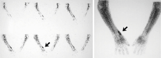
The scintigraphic appearance of de Quervain’s tenosynovitis is noted by hyperaemia on the blood pool phase and focal linear skeletal uptake along the radial aspect of the wrist on the late static views (Fig. 24.2) (Leslie 2006).


Fig. 24.2
De Quervain’s tenosynovitis, blood flow hyperaemia (arrow) and focal linear uptake (arrow) along the radial aspects of the wrist on the late images (Courtesy: Leslie (2006))
24.2.1.2 Proximal Intersection Syndrome
This is an overuse-related inflammatory condition where the tendons of the first dorsal compartment tendons, the abductor pollicis longus (APL) and extensor pollicis brevis (EPB), pass over the tendons of the second dorsal compartment, the extensor carpi radialis longus (ECRL) and extensor carpi radialis brevis (ECRB). Intersection syndrome affects individuals involved in activities requiring repetitive flexion and extension of the wrist and therefore tends to affect sportsmen such as weightlifters, rowers, canoeists, skiers and racket sport players. There is tenderness to palpation and crepitus along the radial aspect of the dorsal distal radius (Dobyns et al. 1978).
In most cases, the clinical presentation is specific; in rare cases it may lead to misdiagnosis or a wide differential diagnosis necessitating imaging for further management.
24.2.1.3 Extensor Carpi Ulnaris Tendinopathy
Extensor carpi ulnaris (ECU) tenovaginitis is the second most common tendinopathy of the wrist (Wood and Dobyns 1986). The extensor carpi ulnaris tendon passes through the sixth dorsal extensor compartment. The tendon may, under appropriate conditions, subluxate or dislocate. ECU tenovaginitis with or without dislocation produces pain and swelling along the dorsal ulnar aspect of athletes who participate in racquet sports, rowing and squash (Kiefhaber and Stern 1992).
Nuclear medicine imaging may prove extremely useful, with the scintigraphic pattern of ECU tenovaginitis which is elongated increased uptake in the blood flow and blood pool phase along the anatomical course of the ECU tendon. Late static images demonstrate increased focal abnormal uptake along the anatomical course.
24.2.1.4 Flexor Carpi Ulnaris Tendinopathy
The flexor carpi ulnaris is a type I tendon; it lacks a synovial tendon sheath; therefore pain associated with this tendon often points to a tendinopathy including a calcific tendinopathy. The condition is associated with repetitive ulnar deviation. Flexor carpi ulnaris (FCU) tendinopathy presents with palmar-ulnar-sided wrist pain and is seen in racquet sport athletes. Examination reveals tenderness along the FCU and pain with wrist flexion (Rettig 2001; Dobyns et al. 1978).
Bone scintigraphy with 99mTc-labelled diphosphonates may demonstrate mild nonspecific uptake, which may help to confirm the clinical suspicion.
24.2.1.5 Flexor Carpi Radialis Tendinopathy
Flexor carpi radialis (FCR) tendinopathy presents with pain in the palmar radial wrist with repetitive wrist flexion. The tendon passes through a narrow fibro-osseous canal at the level of the carpal bones, and it is at greatest risk at this point (Rettig 2001; Dobyns et al. 1978). Tenderness is located over the FCR at its insertion, where it first slips to the trapezium tuberosity then slips to the base of the second and third metacarpals, and pain is reproducible with resisted wrist flexion.
24.2.2 Tendon Tears
Tendon tears may be open, due to lacerations, or closed. Tendon tears more commonly involve the extensor tendons and extensor mechanism of the fingers than the flexor tendons (Clavero et al. 2002). Most open tendon tears are diagnosed and managed clinically and thus not discussed in this chapter. Closed tendon injuries are usually the result of blunt impaction or indirect trauma causing sudden forced stretching of a tendon. Such injuries may also be due to repetitive direct trauma.
24.2.2.1 Mallet Finger
Extensor tendon injury at the distal interphalangeal (DIP) joint is called a mallet finger. It is the most common closed extensor tendon injury in athletes. Sudden, acute, forceful flexion of the extended finger at the DIP joint most frequently accounts for this injury. A classic example is being struck on the fingertip with a ball. Four types of extensor tendon injuries have been described. Type 1, the most common, is extensor tendon avulsion from the distal phalanx with loss of tendon continuity and possibly a small chip avulsion fracture. Type 2 is a tendon and skin laceration at or proximal to the DIP joint. Type 3 is a deep abrasion with skin and tendon loss. Type 4 encompasses fractures of the distal phalanx other than small chip avulsions (Stern and Kastrup 1988). Clinical symptoms are tenderness at the dorsal part of the DIP joint with flexion at the DIP joint and lack of possible active extension of the DIP joint.
Imaging may have added value if an avulsion fracture is present, with bone scintigraphy depicting focal increased accumulation in the area of the extensor tendon at the distal interphalangeal joint. This technique may be useful for detection of avulsion fractures (Huellner et al. 2012; Pin et al. 1988; Akdemir et al. 2004).
24.2.2.2 Jersey Finger
Jersey finger is the most common closed flexor tendon injury which is avulsion of the flexor digitorum profundus (FDP). It most commonly involves the ring finger and occurs in rugby players as the result of grasping an opponent’s clothing (Clavero et al. 2002). Jersey finger results from forceful hyperextension of the DIP joint with the FDP in maximal contraction. Flexor digitorum profundus avulsion is classified into four types, depending on bony involvement and retraction (Leddy and Packer 1977): Type1 is a tendon avulsion without bone fragment with retraction of the flexor digitorum profundus tendon into the palm. Type 2 is a tendon tear without bone fragment with retraction to the proximal interphalangeal (PIP) joint. Type 3 is when an avulsed bone fragment connected to the tendon that is held in place distally by the A4 pulley. Type 4 is type 3 bone avulsion with simultaneous avulsion of the tendon from the bone fragment. Symptoms are tenderness at the volar part of the DIP joint, extension position at the DIP joint and inability to actively flex the DIP joint.
Bone scintigraphy with SPECT/CT and MRI may add new information in patients with clinically nonspecific pain of the ring finger. Bone scintigraphy depicts a characteristic pattern of increased tracer uptake in the region of the flexor digitorum profundus and at the location of the avulsion fracture (Huellner et al. 2012; Pin et al. 1988; Akdemir et al. 2004).
24.2.2.3 Pulley Lesions (Climbers)
The flexor tendons and flexor pulleys are prone to sprains and ruptures. Pulley injuries occur in rock climbers (Peterson and Bancroft 2006). Pulley ruptures related to rock climbing most commonly involve the ring and middle fingers (Klauser et al. 2002). The pulley system refers to a number of retinacular structures that span from side to side across the volar aspect of the fingers. Two types of pulleys, annular pulleys (A1–A5) and cruciform pulleys (C1–C3), are usually identified in the fingers. The annular pulleys are formed by thick arciform fibres, whereas the cruciform pulleys are formed by thin crisscrossing fibres (Peterson and Bancroft 2006; Klauser et al. 2002). Annular and cruciform pulleys extend from the metacarpal heads to the base of the distal phalanges at 8 specific points along the flexor tendon sheath. The A1, A3 and A5 annular pulleys are located over the metacarpophalangeal joint (A1), proximal interphalangeal joint (A3) and distal interphalangeal joint (A5). The A2 and A4 annular pulleys are located over the proximal phalanx (A2) and middle phalanx (A4) (Peterson and Bancroft 2006; Klauser et al. 2002). Injuries typically begin at the A2 pulley, followed by the A3 and A4 pulleys. Pulley injuries can be divided into 3 grades. Grade 1 is sprain of the finger ligaments (collateral ligaments). Grade 2 is partial rupture of the A2 pulley. Grade 3 is complete rupture of the A2 pulley causing bowstringing of the tendon. Climbers present with pain locally at the pulley, pain when squeezing or climbing, active extension deficit and sometimes pain while extending the finger.
Mild nonspecific uptake may be noted on bone scintigraphy, which may help in confirming the clinical suspicion.
24.2.2.4 Boutonnière Injury
Central slip extensor tendon injury may cause a boutonnière injury over time. The boutonnière injury is the least common and perhaps most difficult of the tendon injuries to diagnose. The injury is the result of jamming the finger causing a tear of the extensor tendon attached to the middle phalanx (Aronowitz and Leddy 1998). Patients present with tenderness at the dorsal part of the proximal interphalangeal (PIP) joint (middle phalanx), flexion at the PIP joint and inability to actively extend the PIP joint.
At bone scintigraphy, there are no typical findings. Abnormal (hyperperfusion) blood pool and slightly elevated uptake at the late images in the area of the extensor tendon attached to the middle phalanx can be found; however, this is rather unspecific.
24.3 Nerves
Neuropathies in the upper extremities commonly include acute compression by direct or indirect trauma, chronic irritation related to overuse, pressure related to extreme positions of flexion and extension and repetitive stretching.
24.3.1 Carpal Tunnel Syndrome
Carpal tunnel syndrome (CTS) results from compression of the median nerve, and it is the most common entrapment neuropathy. CTS is common in cyclists, gymnasts, throwers, wheelchair athletes and racquet sport players (Izzi et al. 2001). It is commonly caused by fractures and dislocations or by overuse tenosynovitis of flexor tendons (Bencardino and Rosenberg 2006). The median nerve is entrapped as it passes through the nonyielding carpal tunnel. The athlete has a positive Tinel’s test which is performed by lightly percussing over the nerve to elicit a sensation of tingling, indicating nerve pathology, and a positive Phalen’s test which is elicited by allowing a wrist to fall freely into maximum flexion and maintain the position for 60 seconds or more.
The diagnosis may be made clinically and with the use of nerve conduction studies. Imaging studies may be used to confirm the diagnosis in equivocal cases but, more importantly, may be useful in detecting the underlying cause of median nerve compression.
Levinsohn suggests that a combination of the clinical and appropriate imaging modalities will offer the referring clinician an adequate assessment of the bones and soft tissues of the hands and wrists (Levinsohn 1990). The three-phase bone scan is particularly useful to assess the chronicity of the abnormality. To optimise the value of bone scans of the wrist, the images should be recorded for an extended interval of time. The low concentration of labelled diphosphonate uptake in the wrists of adults, coupled with the complex anatomy, requires high-resolution data and preferable SPECT/CT for definitive diagnosis. The use of three-phase bone scintigraphy assists the referring physician in identifying osseous abnormalities or synovitis as a cause for pain.
24.3.2 Ulnar Neuropathy
Cycling is a particularly common cause of ulnar neuropathy, because the ulnar nerve at the wrist passes through the Guyon’s canal/ulnar canal. The term handlebar palsy refers to a compression syndrome of the deep motor branch of the ulnar nerve and is particularly common in downhill mountain biking (Capitani and Beer 2002). Ulnar neuropathy presents with pain and paraesthesias of the small finger and the ulnar half of the ring finger. Symptoms depend on the location of compression relative to Guyon’s canal.
Bowler’s thumb is a neuropathy of the digital nerve on the ulnar side of the thumb. As the name implies, it is a condition in bowlers who develop compression of the nerve due to keeping their thumbs in the hole of the bowling ball. Imaging is not necessary but MRI may demonstrate perineural fibrosis. No specific scintigraphic pattern can be found; only diffuse uptake may be noted on the blood pool images.
24.3.3 Radial Sensory Neuropathy
The radial nerve is frequently compressed in the distal forearm in its subcutaneous area. The nerve runs subcutaneous between the brachioradialis and extensor carpi radialis longus and is subject to irritation by wristbands and gloves (Izzi et al. 2001). Patients present with pain and decreased sensation over the dorsoradial part of the hand, dorsal thumb and index finger. There is no motor loss. A positive Tinel’s sign may be present with percussion of the nerve. No specific scintigraphic pattern can be found; only diffuse uptake may be noted on the blood pool images.
24.4 Ligaments
Wrist stability is maintained by a series of intrinsic and extrinsic ligamentous structures. The most important intrinsic structures are the scapholunate ligament (SLL), the lunotriquetral ligament (LTL) and the triangular fibrocartilage complex (TFCC) (Lisle et al. 2009). Carpal instability is a continuum of disorders that can result from an acute trauma or from repetitive injury.
Ligament tears are easily visible on CT arthrography or MR (arthrography) due to the leakage of intra-articular contrast medium into the ligaments. Only longstanding ligament lesions are usually associated with changes in the osseous surfaces, i.e. secondary degenerative changes due to advancing instability (Cerezal et al. 2002).
For triangular fibrocartilage complex injuries, MR or CT arthrography is recommended (Zinberg et al. 1988) followed by bone scan if the arthrogram is normal. In the case of suspected bone abnormality, the order of imaging is spot radiographs, tangential radiographs or special radiograph views. This series of studies can then be followed by bone scintigraphy.
In the case of ligamentous injuries, the patient is radiographically evaluated for ligamentous instability. If this exam is normal, then an arthrogram with fluoroscopy is recommended. When the arthrogram with fluoroscopy is unrevealing, bone scintigraphy should be employed.
24.4.1 Scapholunate Injuries
Scapholunate injuries result from a fall on a hand in which the wrist is extended with ulnar deviation. Typical signs are swelling, decreased range of motion and dorsal wrist tenderness in the area of the scaphoid and lunate. A positive scaphoid shift test or Watson’s sign which is done by placing a thumb over the patient’s scaphoid tubercle and applying dorsal pressure indicates a scapholunate tear (Parmelee-Peters and Eathorne 2005). Traumatic tears of the SLL may be partial or full thickness and may involve the central ligament or either one of the bony attachments, more commonly the scaphoid attachment (Connell et al. 2001). Chronic scapholunate ligament tears may result in the presence of ganglion.
The scintigraphic evaluation using three phases will mostly be negative in a partial-thickness tear. The full-thickness tear will demonstrate focal increased activity in the area of the scaphoid and lunate bone.
24.4.2 Lunotriquetral Ligament Tear
Lunotriquetral ligament tears also result from a fall on a dorsiflexed wrist that forces the forearm into pronation (Connell et al. 2001). It is much less common than scapholunate tears. Lunotriquetral tears present with ulnar-sided wrist pain, weakness and possibly clicking. There is tenderness over the area of the lunotriquetral ligament. Dorsal pressure over the pisiform and palmar force on the lunate may produce a painful click.
Bone scintigraphy is less requested for lunotriquetral tears as some of the cases irrespective of whether they have complete or incomplete tear may be normal. In almost half of the cases, there will be focally increased uptake in the first two phases than the 3rd phase. Occasionally diffusely increased uptake at the late phase may be seen throughout the carpus (Huellner et al. 2012; Pin et al. 1988; Akdemir et al. 2004).
24.4.3 Triangular Fibrocartilage Complex Injury
The triangular fibrocartilage complex (TFCC) is composed of the semicircular biconcave fibrocartilage or articular disc called the TFCC, the palmar and dorsal distal radioulnar ligaments, a meniscus homolog and the ulnalunate and ulnatriquetral ligaments (Palmer 1989).
The main function of the TFCC is to act as a strut, which stabilises the distal radioulnar joint while conducting functional pronation and supination activities (Palmer 1989). It acts as a shock absorber when conducting the radial and ulnar deviation activities. The TFCC can sustain injuries in both the medial meniscus disc area and the outer lateral disc areas. This structure is commonly injured with distal radius fractures and wrist sprains. TFCC injuries are more commonly seen in gymnastics, hockey, racquet sports, boxing and pole vaulting (Palmer and Werner 1981).
Palmar’s classification divides TFCC injuries into two types: (1) traumatic (type 1) and (2) degenerative (type 2) (Palmer 1987). Type 1 is further classified into four subtypes based upon the location of the insult. Type 1A is the most common lesion, a horizontal tear in the articular disc adjacent to the sigmoid notch of the distal radius. Type 1B is an avulsion of the TFCC from the ulna. Type 1C is an avulsion of the ulnocarpal ligaments from the carpus. Type 1D is an avulsion from the sigmoid notch of the radius.
Degenerative type 2 tears are believed to result from ulnocarpal impaction. They are divided into five stages: (a) thinning of the TFCC without perforation, (b) thinning of the disc with chondromalacia of the ulnar head or lunate, (c) disc perforation with chondromalacia, (d) disc perforation with chondromalacia and lunotriquetral ligament tear and (e) disc perforation with chondromalacia, lunotriquetral tear and ulnocarpal arthritis. Central perforations are usually attributed to degenerative processes (Palmer 1989).Triangular fibrocartilage complex perforations increase with age. Ulnar-sided wrist pain with tenderness on palpation of the TFCC, with or without distal radioulnar joint instability, is indicative of TFCC injury.
24.4.4 Distal Radioulnar Joint Instability
The distal radioulnar joint (DRUJ) is located between the distal radius and the head of the ulnar. Five structures are important in ensuring the stability of the DRUJ: (1) the TFCC, (2) the ulnocarpal ligament complex, (3) the infratendinous extensor retinaculum (i.e. the ECU tendon sheath), (4) the pronator quadratus muscle and (5) the interosseous membrane (Garcia-Elias and Dobyns 1998). The DRUJ has intimate and overlapping structures with TFCC; hence, injury is caused by similar mechanisms. Like TFCC, it presents with ulnar-sided wrist pain. The most common cause of DRUJ instability is a result of TFCC injury since TFCC is significant to DRUJ stability. Other causes of instability are distal radius fractures or disruption of any of the distal radioulnar ligaments or interosseous membrane (Parmelee-Peters and Eathorne 2005).
Currently there is no evidence on the role of bone scintigraphy in DRUJ. An indirect role of scintigraphy is that of being sensitive in severe/complete TFCC.
24.4.5 Skier’s (Gamekeeper’s) Thumb
A tear of the ulnar collateral ligament (UCL) at the MCP joint of the thumb on the side of the webspace is called a skier’s thumb (Spaeth et al. 1993). This injury is caused by violent abduction of the thumb. This injury was historically named gamekeeper’s thumb because of its frequency in the gamekeepers of the large hunting estates in England. The injury is most commonly seen in football players and skiers. The two classifications of this injury are (1) incomplete, or proper collateral ligament injury with accessory collateral preserved, and (2) complete, in which the ligament rupture is complete and the distal end may displace superficial and proximal to the adductor aponeurosis. This condition is referred to as a Stener’s lesion (Stener 1962). In patients with complete ruptures, the incidence of adductor aponeurosis interposition and ligament displacement is controversial but has been reported in as many as 80 % of cases (Bowers and Hurst 1977).
At bone scintigraphy, hyperaemia at the blood pool phase and moderate increased osseous uptake at the late phase will be seen on the UCL at the MCP joint of the thumb on the side of the webspace. These findings are better seen on SPECT/CT (Huellner et al. 2012).
24.5 Fractures
Bone scintigraphic evaluation of wrist and hand injuries is often a very helpful procedure when fractures are suspected, especially with nondiagnostic radiographs. These may include fractures of the distal radius and ulna and the carpal or metacarpal and phalangeal regions. SPECT/CT may help to improve the sensitivity and specificity by better resolution and exact localisation of the increased uptake.
In a study to evaluate the role of bone scintigraphy in patients with suspected carpal fracture and normal or suspicious radiographs following carpal injury, a three-phase 99mTc-MDP scan was performed on 32 patients with negative radiographs but clinically suspected fracture two weeks after the trauma. Twelve (38 %) patients had a normal scan excluding fracture. Twelve patients had a single fracture. Multifocal fracture was present in 8 (25 %) patients. Eight patients showed scaphoid fractures: three showed single scaphoid fracture, and the other five patients revealed accompanying fractures (Weber 2011). Distal radius fractures and carpal bone fractures other than scaphoid were both observed in 12 patients. These were eleven fractures of distal radius; three fractures of pisiform; two fractures of hamate; and single fractures of lunate, trapezium and triquetrum. In one patient there was a fracture of a first metacarpal bone. In patients with suspected carpal bone fracture and normal or suspicious radiographs, bone scintigraphy can be used as a reliable method to confirm or exclude the presence of a scaphoid fracture and to detect clinically unsuspected fractures of distal radius and other carpal bones (Akdemir et al. 2004). In another study involving 87 patients, the sensitivity and negative predictive value for carpal fractures were 97 and 98 %, respectively, highlighting the benefit of nuclear medicine imaging techniques in the detection of occult carpal fractures, thereby reducing the risks of complications such as pseudoarthritis (Querellou et al. 2009).
24.5.1 Distal Radius Fracture
A fracture of the distal radius is the most common forearm fracture. It is usually caused by a fall onto an outstretched hand (FOOSH). It can also result from direct impact or axial forces. The classification of these fractures can be based on distal radial angulation and displacement, intra-articular or extra-articular involvement and associated anomalies of the ulnar or carpal bones (Rettig and Trusler 1999).
Stay updated, free articles. Join our Telegram channel

Full access? Get Clinical Tree


