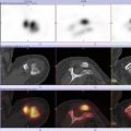
Fig. 15.1
A 52-year-old amateur cyclist was admitted to the hospital after a cycling accident while cycling 35 km/h. He presented with pain between the scapulae, in the lower neck, and in his thorax. The initially performed CT only showed a fracture of the left clavicle. Due to the continuing pain complaints, a bone scintigraphy with SPECT-CT was performed. This revealed a rib fracture of the 3rd rib on the right side, a fracture of the right processus spinosus of the 1st thoracic vertebra, and a fracture of the processus spinosus of the 4th rib (Courtesy of H. Balink, Department of nuclear medicine, Medical Centre Leeuwarden)
15.2.1.2 Acute Rib Fractures
Diagnosis of rib fractures may be difficult. Identification of rib fractures by plain radiography can be challenging, but plain radiographs are very helpful in detecting additional disease such as hemothorax or pneumothorax. After the acute phase, bone scintigraphy with SPECT/CT can be performed to examine the presence of rib fractures (Erhan et al. 2001). Nevertheless, not only plain radiographs are limited in detection of rib fractures, but this also applies to CT. In a recent study involving 130 patients suffering from blunt chest trauma, 52 rib fractures were missed on CT reconstructions (Cho et al. 2012). In these cases the combination of a SPECT and CT will likely give a higher sensitivity and specificity for the detection of rib fractures. Moreover, bone bruising can be distinguished from fractures using the CT and thereby increases the diagnostic advantage of bone SPECT-CT for the clinician.
15.2.1.3 Rib Stress Fractures
A stress fracture can be defined as a partial or complete bone fracture resulting from repetitive stress, stress milder than required in order to fracture the bone in a single loading. Stress fractures occur when the remodeling and repair capabilities of the bone are exceeded by the continuously occurring microfractures during repetitive exercise (Bennell et al. 1996).
The most common stress fracture occurs in the first rib. Nevertheless, many fractured first ribs remain asymptomatic and are coincidentally found. In sports involving overhead activity, the repetitive contractions of the scalene muscles produce bending forces which may finally result in a fracture (Connolly and Connolly 2004). Pain can be located in the shoulder, be linked to inspiration, or be tenderness in the neck. In these cases chest radiographs are of limited help, and bone scintigraphy, CT, and MRI are more sensitive. Generally, MRI has the advantage of being able to exclude other pathologies without radiation risk, whereas bone scintigraphy is better in excluding osseous pathology (Dobrindt et al. 2012).
In golf players and elite rowers, fractures of other ribs are more common. These fractures are as well often difficult to ascertain on plain radiographs, and for the diagnosis of these fractures, bone scintigraphy and possibly NaF PET are required (Maffulli and Pintore 1990; Karlson 1998; Gregory et al. 2002). However, NaF PET is likely to have a superior sensitivity in the detection of (impending) stress fractures compared to bone scintigraphy, but its value has to be ascertained (Drubach et al. 2010; Li et al. 2011; Iagaru et al. 2012).
15.2.2 Sternum Fractures
Only 0.5 % of all sternum fractures are sports-related stress fractures, usually occurring in sports requiring extreme upper body stresses, e.g., wrestling, strenuous abdominal muscle training, and even golfing (Keating 1987; Barbaix 1996; Gregory et al. 2002). The subsequent pain is generally gradual or sudden in onset, is located in the anterior chest, and increases with deep breathing. For the diagnosis of sternal fractures, ultrasound, plain radiographs, or CT can be applied. For the detection of sternal fractures in the early phase, both ultrasound and bone scintigraphy can be used (Erhan et al. 2001; Jin et al. 2006). However, as with rib fractures, NaF PET may be more sensitive than bone scintigraphy (Drubach et al. 2010; Li et al. 2011).
15.2.3 Costochondritis
Costochondritis is a poorly understood condition (Proulx and Zryd 2009), characterized by tenderness and pain on the costochondral and costosternal joints, though without swelling of the involved joints. Costochondritis has a higher prevalence in both athletic young females as in young females in the general population. Regarding sports, this syndrome is most common in young female rowing athletes (Grindstaff et al. 2010). Bone scintigraphy in patients with costochondritis generally shows an increased uptake in costochondral joints, but this finding is not specific for this condition (Mendelson et al. 1997). Nonetheless, bone scintigraphy may have additional value in combination with CT and plain radiographs.
15.3 Lung Injuries
15.3.1 Pneumothorax and Tension Pneumothorax
Pneumothorax is a common complication following blunt chest wall trauma. Generally plain PA chest radiographs are acquired for the diagnosis of pneumothorax. Traditionally, nuclear medicine is not used in the diagnosis of pneumothorax (Omar et al. 2011).
However, ventilation perfusion scintigraphy can be used to examine impairment of regional or overall lung function. In patients diagnosed with pneumothorax, impaired ventilation and perfusion was found in the apical regions of the lungs where the pneumothorax had occurred (Bense et al. 1986). In a published case report, lung ventilation and perfusion scintigraphy contributed to the diagnosis of an unsuspected pneumothorax (Lee et al. 1984). Whereas lung perfusion scintigraphy, with or without SPECT/CT, has no place in routine evaluation of pneumothorax, it might nevertheless be of use in localizing the air leaks responsible for pneumothorax (Ceulemans et al. 2012).
15.4 Conclusion
Due to the low incidence of traumatic sports and stress injuries to the thorax, only scarce research data exists about the use of nuclear medicine imaging techniques in this part of the musculoskeletal system. However, the use of NaF PET might be beneficial for the detection of traumatic ossal injuries to the thorax. Further research and experience with this technique for this application is warranted.
References
2011-187 verdict of dutch disciplinary law august 7th 2012 (2012) In ’s-Gravenhage RTvdGt (ed), (ed), Vol. pp. Overheid.nl, ’s-Gravenhage
Barbaix EJ (1996) Stress fracture of the sternum in a golf player. Int J Sports Med 17:303–304CrossRefPubMed
Related posts:


Stay updated, free articles. Join our Telegram channel

Full access? Get Clinical Tree


