Presentation and Presenting Images
( ▶ Fig. 51.1, ▶ Fig. 51.2, ▶ Fig. 51.3, ▶ Fig. 51.4)
A 74-year-old female presents for screening mammography.
51.2 Key Images
( ▶ Fig. 51.5, ▶ Fig. 51.6, ▶ Fig. 51.7, ▶ Fig. 51.8)
51.2.1 Breast Tissue Density
There are scattered areas of fibroglandular density.
51.2.2 Imaging Findings
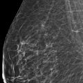
There are two lesions identified on the screening mammography examination. At the 11 o’clock location in the posterior depth, there appears to be a mass with a partially convex margin ( ▶ Fig. 51.5 and ▶ Fig. 51.6). Additionally, lateral in the right breast, there is architectural distortion in the middle depth only seen on the craniocaudal (CC) view ( ▶ Fig. 51.1). This is better appreciated on the CC synthetic mammogram ( ▶ Fig. 51.8) and tomosynthesis images ( ▶ Fig. 51.7).
51.3 BI-RADS Classification and Action
Category 0: Mammography: Incomplete. Need additional imaging evaluation and/or prior mammograms for comparison.
51.4 Diagnostic Images
( ▶ Fig. 51.9, ▶ Fig. 51.10, ▶ Fig. 51.11, ▶ Fig. 51.12, ▶ Fig. 51.13, ▶ Fig. 51.14, ▶ Fig. 51.15, ▶ Fig. 51.16, ▶ Fig. 51.17)
51.4.1 Imaging Findings
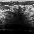
The additional diagnostic imaging confirms the 1.8 × 1.4 cm irregular mass with indistinct calcifications at the 10 o’clock location in the posterior depth ( ▶ Fig. 51.9 and ▶ Fig. 51.10). Ultrasound reveals a corresponding 1.4 × 1.3 × 1.0 cm irregular mass with indistinct and angular margins and posterior acoustic shadowing at the 10 o’clock location, 8 cm from the nipple ( ▶ Fig. 51.12). With further review of the initial digital breast tomosynthesis (DBT) images ( ▶ Fig. 51.11), the architectural distortion appears to have a correlate on the mediolateral oblique (MLO) view, at the 9 o’clock location. Ultrasound of this region reveals a 1.2 × 0.8 × 0.7 cm irregular mass with indistinct margins, 5 cm from the nipple ( ▶ Fig. 51.13). Similar to the 10 o’clock lesion, this mass also has posterior acoustic shadowing. These lesions occupy the same quadrant of the breast with the 9 o’clock mass anterior and medial to the 10 o’clock mass and separated by 4 cm ( ▶ Fig. 51.14). Both lesions were biopsied under ultrasound guidance. The 10 o’clock lesion was marked with a coil biopsy clip and the 9 o’clock lesion was marked with a ribbon clip ( ▶ Fig. 51.15 and ▶ Fig. 51.16). The lesions and their respective biopsy clips can be seen in this lumpectomy specimen ( ▶ Fig. 51.17).
51.5 BI-RADS Classification and Action
Category 5: Highly suggestive of malignancy
51.6 Differential Diagnosis
Invasive lobular carcinoma [ILC]: Both of these lesions were found to be grade 1 ILC.. It is important to be aware of the possibility of additional lesions in the breast when an initial lesion is detected.
Invasive ductal carcinoma [IDC]: The presentation and imaging characteristics of these lesions would be also be consistent with IDC.
Fibrosis: Fibrosis can mimic carcinoma. To be concordant on image-guided biopsy, the size of the lesion on imaging should closely match the size on histopathology. Both of these lesions are highly suspicious. If there is uncertainty in the radiology–pathology concordance, an excision would be warranted.
51.7 Essential Facts
The nature of ILC, in its tendency to produce little desmoplastic reaction and its single file growth pattern within the breast tissue, can make it difficult to detect both clinically and by imaging.
ILC has a variable appearance; it may present as a focal asymmetry, an architectural distortion, a mass, or with no mammographic findings (occult).
The unmasking benefit of DBT may prove beneficial to the detection of the subtle lesions of ILC. Early studies suggest that there is a higher proportion of ILC detected in the studies the include DBT compared to those that use full-field digital mammography (FFDM) alone.
51.8 Management and Digital Breast Tomosynthesis Principles
10 to 30% of breast cancers are missed on conventional mammography.
The majority of FFDM occult lesions that were identified on DBT were in dense breasts.
It is important not to rush mammographic interpretation and settle for the first lesion that is detected (“satisfaction of search”).
DBT is very good at detecting architectural distortion. However, not all architectural distortion is cancer. Radial scars and atypical ductal hyperplasia (ADH) are among the frequent sources of false-positive biopsies on DBT.
51.9 Further Reading
[1] Majid AS, de Paredes ES, Doherty RD, Sharma NR, Salvador X. Missed breast carcinoma: pitfalls and pearls. Radiographics. 2003; 23(4): 881‐895 PubMed
[2] Ray KM, Turner E, Sickles EA, Joe BN. Suspicious Findings at Digital Breast Tomosynthesis Occult to Conventional Digital Mammography: Imaging Features and Pathology Findings. Breast J. 2015; 21(5): 538‐542 PubMed

Fig. 51.1 Right craniocaudal (RCC) mammogram.
Stay updated, free articles. Join our Telegram channel

Full access? Get Clinical Tree


