Summary of Key Points
- •
Obesity is epidemic and a significant public health problem.
- •
Obese women are at increased risk of maternal complications of pregnancy compared to women of normal weight.
- •
Fetuses of obese gravidas are at increased risk of malformations compared to those of normal weight women.
- •
Ultrasound imaging of obese patients is hampered by acoustic noise, artifacts, and thicker anterior abdominal wall, all of which degrade the image.
- •
Although ultrasound examinations of obese gravidas often are repeated in order to obtain adequate images, fetal anatomic evaluation is frequently suboptimal despite several attempts.
- •
There are several ways to optimize the image quality while scanning obese patients.
Over 60% of the population of the United States is considered overweight, and more than one third is obese, making obesity a significant public health problem. In 2005, the World Health Organization estimated that 1.6 billion adults worldwide were overweight and 400 million were frankly obese (body mass index [BMI] ≥ 30). This worldwide epidemic shows no sign of abating and continues to increase, especially in the United States. A growing body of evidence shows that obese patients have a higher risk of a multitude of health problems, including heart disease, type 2 diabetes, hypertension, stroke, “metabolic syndrome,” arthritis, cancer, respiratory problems, sleep apnea, pregnancy complications, and more ( Table 25-1 ). The National Institutes of Health and the World Health Organization define obesity according to the BMI (in kg/m 2 ). A BMI of 18 to 24.9 is normal, and a BMI of 25 to 29.9 is overweight. Classes I, II, and III obesity are defined as BMIs of 30 to 34.9, 35 to 39.9, and 40 or higher, respectively.
| Maternal Risks | Fetal Risks |
|---|---|
| Cardiovascular disease | Congenital anomalies |
| Hypertension | Lower detection of fetal anomalies |
| Recurrent miscarriage | Macrosomia |
| Gestational diabetes | Inaccurate fetal weight estimate |
| Preeclampsia | Preterm birth |
| Cesarean delivery | Stillbirth and neonatal death |
| Postpartum hemorrhage | |
| Wound infection | |
| Thromboembolism | |
| Maternal death |
Obese women have an increased risk for adverse pregnancy outcomes that impact both the mother and the fetus. Sebire and associates described 287,213 patients, of which 39% were obese, and reported that several maternal complications increased with the degree of obesity. Table 25-1 lists the most common maternal and fetal complications associated with maternal obesity. In addition, an increased risk of fetal malformations has been associated with obesity, and fetal anomalies such as neural tube defects, cardiac anomalies, anal atresia, and limb reduction abnormalities are reported to be significantly more common with obese patients compared to the normal weight population ( Table 25-2 ).
| Anomaly | OR (95% CI) |
|---|---|
| Anal atresia | 1.48 (1.12-1.97) |
| Cardiac defects | 1.30 (1.12-1.51) |
| Hydrocephalus | 1.68 (1.19-2.36) |
| Limb reduction | 1.34 (1.03-1.73) |
| Neural tube defect | 1.87 (1.62-2.15) |
Uhden and colleagues reported that the relative risk of heart defects is 2.04 in fetuses of obese compared to normal weight women. Watkins and coworkers also demonstrated that obese compared to average weight women have an elevated risk of fetal anomalies such as spina bifida (odds ratio [OR] 3.5; 95% confidence interval [CI] 1.2-10.3), omphalocele (OR 3.3; 95% CI 1.0-10.3), and heart defects (OR 2.0; 95% CI 1.2-3.4), as well as multiple anomalies (OR 2.0; 95% CI 1.0-3.8). In a study evaluating risk factors for fetal malformations, Waller and associates showed that patients who had infants with spina bifida, heart defects, anorectal atresia, hypospadias, limb reduction defects, diaphragmatic hernia, and omphalocele were significantly more likely to be obese, with ORs between 1.33 and 2.10. A meta-analysis of 12 studies demonstrated that the OR for having a fetus with a neural tube defect was 1.22, 1.70, and 3.11 for overweight, obese, and severely obese patients compared to normal weight patients, indicating that the risk rises with increasing obesity.
Obese gravidas require more medical resources than normal weight patients and often present difficult technical problems for the medical personnel providing their care. High BMI patients have been reported to require more interventions and care of all types, including obstetric ultrasound examinations, medications, telephone calls to providers, and prenatal visits with physicians compared to women of normal weight.
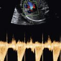
In addition to requiring more numerous ultrasound examinations, the examinations required to detect fetal anomalies and pregnancy complications are often more technically challenging in obese compared to thinner patients. The quality of the ultrasound examination done on an obese patient is technically inferior when compared to the same scan done on slimmer patients owing to the distance traversed by the sound beam and the density of adipose tissue. Hence, the quality of the ultrasound image relates inversely to the distance that the beam must travel to reach the fetus. The ultrasound beam is absorbed, dispersed, and attenuated in adipose tissue, limiting the image quality in evaluation of the fetus. The ultrasound image is further degraded with each centimeter that it penetrates through the body to reach the fetus. As a result, the most obese patients who are at the highest risk of maternal and fetal complications also have the poorest quality imaging by ultrasound. Over time, advances in ultrasound technology have resulted in improved image quality even in the more challenging patients. Preprocessing and postprocessing filters, speckle-reduction filters, compound imaging, and tissue harmonic imaging have all contributed to improved image quality. Despite these new tools, the ultrasound image in obese gravidas often remains fuzzy and noisy and suffers from excessive backscatter and artifacts ( Fig. 25-1 ).

A number of studies have compared the ability to evaluate the fetus and detect fetal anomalies in obese versus normal weight women. Stothard and colleagues showed that although the risk of many fetal anomalies is significantly higher in obese compared with normal weight patients, a fetal anatomic survey was not completed at the time of the first ultrasound detailed scan in more than 50% of obese women. Dashe and associates evaluated the quality of second trimester ultrasound studies done at 18 to 24 weeks on a cohort of 10,112 patients, 34% of whom were overweight and 26% of whom were obese. The following 10 anatomic views were evaluated for adequacy: cerebral ventricles, posterior fossa, midline face (profile), four-chamber view of the heart, spine, ventral wall, cord vessels, stomach, kidneys, and bladder. Table 25-3 lists the relationship between successful scans and obesity class, showing a steady decline in scan quality with increasing BMI. Maxwell and coworkers compared patients with a BMI of 30 or higher (average 35.7) with those having a BMI lower than 25 and found that 26% of the anatomic surveys in obese women were incomplete compared to 2.5% of those in women with a normal BMI. Uhden and colleagues also reported that the percentage of poor ultrasound images rose from 6.4% to 17.4% when comparing normal weight to obese patients, whereas the prevalence of heart defects was higher (relative risk = 2.04) in obese and overweight women when compared to normal weight patients.
Related posts:
 Ultrasound Evaluation of the Fetal Central Nervous System
 Measurements Frequently Used to Estimate Gestational Age and Fetal Biometry
 Role of Doppler Sonography in Obstetrics
Medications and Reported Associated Malformations
Role of Doppler Sonography in Obstetrics
Medications and Reported Associated Malformations
 Role of Sonography in Gynecologic Interventions
 Ultrasound Features of Fetal Syndromes
Stay updated, free articles. Join our Telegram channel

Full access? Get Clinical Tree


