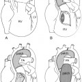
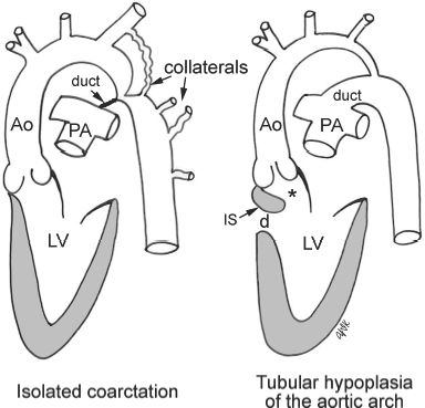
23 Obstructive Lesions of the Aortic Arch Fig. 23.1 Coarctation and tubular hypoplasia of the aortic arch. Tubular hypoplasia of the aortic arch is usually associated with left ventricular outflow flow tract obstruction (asterisk) due to posterior malalignment of the infundibular septum (IS). Ao, ascending aorta; d, ventricular septal defect; LV, left ventricle; PA, pulmonary artery.
 Obstructive lesions of the aortic arch include the following varieties:
 Coarctation and Tubular Hypoplasia of the Aortic Arch
Definition and Classification
 Congenital stenosis of the thoracic aorta of variable severity
 Severe forms leave only a small residual lumen, with mild forms producing only minimal narrowing.
 The terms preductal and postductal as well as infantile and adult type are misleading.
 Coarctation versus tubular hypoplasia (Fig. 23.1)
 Simple aortic coarctation refers to discrete coarctation in the absence of other intracardiac lesions, with the exception of a bicuspid aortic valve.
 Localized ridge-like infolding almost always in a juxtaductal position
 Diffuse with a long hypoplastic segment proximal to the ductus arteriosus
 Complex aortic coarctation refers to coarctation in the presence of other intracardiac anomalies and almost always includes a patent ductus arteriosus. The right panel of Fig. 23.1 shows an example with a posterior malalignment type of ventricular septal defect causing subaortic stenosis.
 Other associations include Berry aneurysms of the circle of Willis.
 Association with Turner syndrome
Pathophysiology
 Simple aortic coarctation is not associated with cardiac defects, and collateral circulation develops gradually over time.
 Complex aortic coarctation is almost always associated with conditions that decrease antegrade flow through the ascending aorta and increase right-to-left shunting across the ductus arteriosus in utero, including ventricular septal defect and left-sided obstructive lesion, such as subaortic stenosis and mitral valve anomalies (Fig. 23.1, right panel).
 Discrete juxtaductal aortic narrowing results in systolic overloading and hypertrophy of the left ventricle.
 In weeks to months as the left ventricle hypertrophies and dilates, the ejection fraction improves and the cardiac output reaches the high normal range.
 At rest left ventricular pressures are normal or mildly increased, but they increase to abnormal levels when afterload is increased with exercise.
 Progressive development of a collateral circulation between the proximal high pressure and the distal low pressure aorta below the coarctation
 Usual vessels developing a collateral circulation are from the subclavian arteries and arteries around the scapula, internal mammary arteries, and the intercostal arteries.
 Blood flow through renal, splanchnic, and leg arteries is normal at rest, but may not increase appropriately with exercise.
 Complex and long segment hypoplasia results in systolic overloading of the left ventricle as well as pulmonary hypertension.
 Right ventricular systolic overloading, right ventricular hypertrophy, and elevated right-sided pressures develop.
 Good collateral circulation has not yet developed, with blood being delivered to the descending aorta from a patent ductus arteriosus.
 Ductal lumen provides additional space to the narrowed aorta.
 Hemodynamic deterioration occurs rapidly at the time of ductal closure.
 Cardiac decompensation, cardiac enlargement, and congestive heart failure develop.
Clinical Manifestations
Stay updated, free articles. Join our Telegram channel

Full access? Get Clinical Tree


