
Fig. 12.1
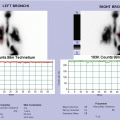
(a, b) Contrast-enhanced CT. Axial (a) and MPR coronal (b) images showed a large retroperitoneal mass, appearing not homogeneously hypodense for the presence of multiple nonenhancing necrotic areas and tiny calcifications in the contest. The lesion, growing behind the aorta, encased left renal artery and displaced left adrenal gland and left kidney; abdominal aorta and inferior cava were pervious, although stretched, flattened, and displaced anteriorly, as well as the splenic vessels, pancreatic tail, and left renal vein. (c–e) MIBG scintigraphy (static views, c) showed an area of intense radiotracer uptake in the left abdomen, crossing the median line; no other areas of pathological uptake of radiotracer were evident. SPET-CT fused images (d, e) better defined that the area of uptake corresponded to the mass detected by CT
The patient was enrolled in LINES protocol stage L2 <18 months NCA genomic profile with life-threatening symptoms (arterial hypertension).
The patient received chemotherapy according to the protocol, and because of the persistence of arterial hypertension, the chemotherapy was prolonged for a total two courses of carboplatin/etoposide plus two courses of cyclophosphamide/vincristine/doxorubicin. The patient underwent surgery at the end of chemotherapy.
12.2.2 Case 12.2 MIBG Scintigraphy in Diagnostic Workup of Abdominal Mass: MIBG – Nonavid Lesion
A 2-year-old baby referred to our hospital for fever and limping; an ultrasonography revealed a median retroperitoneal lesion; therefore, a computed tomography (CT) was performed, confirming the presence of a large retroperitoneal mass, extending from celiac artery to mesenteric artery, suspected for neuroblastoma (Fig. 12.2a–c) Therefore, a MIBG scintigraphy was scheduled in order to verify if the lesion shows MIBG uptake, confirming the diagnosis. MIBG scintigraphy (Fig. 12.2d–e) did not show any area of pathological uptake of radiotracer; in particular, the abdominal lesion detected by CT appears as nonavid MIBG.


Fig. 12.2
(a–c) Contrast-enhanced CT axial (a), MPR coronal (b), and sagittal (c) images showed a large retroperitoneal mass, appearing polylobulated with well-defined borders, about 55 × 65 × 45 mm in size. The mass grew on the midline, between celiac artery and superior mesentery artery that were stretched and displaced, respectively, upper and lower in the abdomen. The lesion displaced inferior cava vein, pancreatic head and body, and portal vein, without signs of infiltration. There were also multiple enlarged nodes in mesenteric fat. (d, e) MIBG scintigraphy (static views, d) did not show any area of pathological uptake of radiotracer; in particular, the abdominal lesion detected by CT appeared as nonavid of MIBG. SPET-CT fused images (e) confirmed this finding
Subsequently, a biopsy of the lesion was performed, and histological examination showed a ganglioneuroma. As in this case, MIBG uptake and proliferative activity are strictly correlated, because MIBG reflects metabolic activity of tumor derived from neural crest; in fact, tumors with a high proliferative activity show higher MIBG uptake than lesions with lesser proliferation, such as ganglioneuromas or ganglioneuroblastoma, as reported in literature.
12.2.3 Case 12.3 Bone Scan in Paravertebral Mass: Ewing’s Sarcoma
An 8-year-old girl referred to our hospital for dorsal pain and swelling; an ultrasonography revealed a right paravertebral lesion; therefore, a CT was performed, showing a large paravertebral mass, located above the right kidney, involving posterior segments of the ribs (Fig. 12.3a, b); histological examination indicated Ewing’s sarcoma/PNET. The girl underwent a whole-body bone scan as part of diagnostic workup. Scintigraphy shows diffuse radiotracer uptake by right XII rib, without other pathological findings in the skeleton (Fig. 12.3c), and the girl was scheduled for therapy, adequate for tumor stage.

Fig. 12.3
(a, b) Contrast-enhanced CT axial (a, b) showed a large right suprarenal mass (about 8 × 8 × 15 cm in size), extending above and below the diaphragm with encasement of the back ribs and of the regional muscle tissue. The lesion, appearing nonhomogeneous after contrast agent administration, dislocated the right kidney inferiorly; liver and inferior cava vein were anteriorly compressed. (c) 99mTc-MDP whole-body scan; right posterior oblique (RPO) and left posterior oblique (LPO) views of thorax: a pathological, intense, and diffuse radiotracer uptake is evident in right XII rib; no other pathological findings are evident in the skeleton
12.2.4 Case 12.4 Bone Scan in Thoracic Wall Sarcoma: Evidence of Bone Metastases
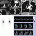
A 15-year-old girl referred to our institution for swelling in correspondence to one of the last ribs of right hemithorax; during diagnostic workup, the girl underwent chest radiography, CT scan, and MRI, and a mass involving right X rib was detected (Fig. 12.4a–c). Histological examination revealed Ewing’s sarcoma/PNET; therefore, a whole-body bone scan was performed for staging purpose (Fig. 12.4d, e). On the basis of scintigraphic results, a diagnosis of metastatic Ewing’s sarcoma was made, and the girl began chemotherapeutic treatment.


Fig. 12.4
(a): Anterior–posterior (AP) plain film of the chest and the abdomen: structural alteration of the IX and X right ribs (black arrows) with soft-tissue swelling (white arrows). (b): CT scan in the coronal, sagittal, and axial planes well highlights the costal osteolysis surrounded by inhomogeneous mass of soft tissue that displaces to the left of the liver and the right kidney. (c) MRI in the coronal and axial planes T1 fat/sat after contrast medium shows the mass of soft tissue with multiple hypointense areas in the context surrounded by peripheral ring enhancement related to necrotic areas. (d, e) 99mTc-MDP whole-body scan (d): a wide area of pathological, intense radiotracer uptake involving the anterolateral part of right X rib corresponding to the mass detected by CT; two small areas of pathological focal uptake are also evident in the posterior part of right XI rib and in the spinous process of one of the first cervical vertebrae (better defined in lateral views of skull, (e), respectively. A right posterior view of thorax (e) with arms up is also performed in order to define the focal uptake evident in inferior angle of right scapula, and it shows that it is due to an overlap of rib and scapula
12.2.5 Case 12.5 Bone Scan in Renal Sarcoma
Staging bone scan in a 14-month-old baby with clear cell renal sarcoma, with lymph node and lung metastasis.
Multiple static views of skeletal segments are acquired, instead of the usual whole-body modality, due to the poor cooperation of the patient (Fig. 12.5).

Fig. 12.5
99mTc-MDP scintigraphy, multiple views of the skeleton: physiological distribution of radiotracer uptake and no evidence of pathological uptake in the skeleton
12.2.6 Case 12.6 Bone Scan in Pelvic Mass: Staging of Malignant Germinoma
A 9-year-old girl referred to our institution for vaginal bleeding; ultrasonography detected a pelvic mass; aFP and bHCG were very high; CT scan confirmed a large pelvic neoplasia involving right ovary and shows also a lung metastasis. Histological examination of the mass revealed a malignant germinoma, and a whole-body bone scan was performed to assess secondary bone involvement (Fig. 12.6).

Fig. 12.6
99mTc-MDP whole-body scan (a) shows physiological distribution of radiotracer uptake and no evidence of pathological uptake in the skeleton. Stasis of radioactive urine is evident in both kidneys and ureters (especially in right one), due to secondary hydroureteronephrosis. A SPECT of the pelvis is also performed, and SPECT-CT (b) manual fused image confirms the absence of pathological uptake in the pelvis and lumbar and sacral spine
12.2.7 Case 12.7 Bone Scan in Rhabdomyosarcoma
A 12-year-old boy referred to our hospital for swelling in right axilla; the boy underwent an ultrasonography and a CT scan, showing a neoplasia in pectoral muscles with involvement of axillary lymph nodes (histological examination: alveolar rhabdomyosarcoma). A whole-body bone scan was also performed for staging purpose (Fig. 12.7a, b); whole-body scan (a) showed a focal area of intense radiotracer uptake (better defined in lateral views of skull, (b) in superior part of left orbit; another small area of focal uptake is evident in the left posterior temporal bone. Furthermore, linear and diffuse radiotracer uptake is detectable in the right forearm, possibly due to the previous traumatic accident referred during clinical interview.


Fig. 12.7
(a, b) Whole-body bone scan (a) shows a focal area of intense radiotracer uptake (better defined in lateral views of skull, (b) in superior part of left orbit; another small area of focal uptake is evident in left posterior temporal bone. Furthermore, linear and diffuse radiotracer uptake is detectable in right forearm, possibly due to the previous traumatic accident referred during clinical interview. (c, d) CT scan in axial, coronal, and sagittal planes with a soft tissue “window” (c); CT scan in axial, coronal, and sagittal planes with “bone window”(d). Small lenticular mass just less dense than the bone starting from the left orbital roof which protrudes inside the orbit contacting the eyeball and the other intraorbital structures. (e, f) Whole-body bone scan (e), after chemotherapy and radiotherapy, shows persistence of a faint focal area of radiotracer uptake in superior part of left orbita (better defined in lateral views of skull, f). (g) Whole-body scan (g) reveals multiple areas of pathological uptake of radiotracer in the whole skeleton
A CT scan (Fig. 12.7c, d) was performed, showing a hyperdense tissue, which extended to the left orbital cavity, starting from inferior border of the orbit roof.
Lesion was surgically removed, and histological examination revealed a lacrimal gland with MALT-tissue activation; after surgery, the boy went on with chemotherapeutic and radiotherapeutic treatment; 99mTc-MDP whole-body scan (Fig. 12.7e), after chemotherapy and radiotherapy, showed persistence of a faint focal area of radiotracer uptake in the superior part of left orbita (better defined in lateral views of skull (Fig. 12.7f). One year after stop therapy, the boy underwent a restaging workup for relapse of disease, including a 99mTc-MDP whole-body scan (Fig. 12.7g) that revealed multiple areas of pathological uptake of radiotracer in the whole skeleton.
12.2.8 Case 12.8 Bone Scan in Large Chest Mass with Limited Bone Involvement
A 15-year-old girl referred to our hospital for diagnostic assessment of a thoracic mass, previously investigated in another center. Chest radiography (Fig. 12.8a) detected a diffuse opacity of the whole left lung field, and subsequent CT scan (Fig. 12.8b) showed a left lung mass eroding a rib, with pleural effusion. Histological examination of the mass showed Ewing’s sarcoma, and a bone scintigraphy was scheduled for staging purpose. 99mTc-MDP whole-body scan (Fig. 12.8c, d) shows a pathological radiotracer uptake in the anterolateral part of a rib of the left hemithorax (probably IV rib) and no other pathological findings.


Fig. 12.8


(a) Frontal chest X-ray. Complete opacification of the left hemithorax with moderate displacement of the trachea, mediastinal structures, and the central venous catheter to the right. (b) CT scan before and after contrast medium. A big mass with multiple hypodense areas with small peripheral enhancement after contrast medium for prevalence of necrotic areas. Dislocation of the mediastinal structures toward the right side. (c, d) 99mTc-MDP whole-body scan (c) shows a pathological radiotracer uptake in anterolateral part of a rib of left hemithorax (probably IV rib), better defined in RAO and LAO views (d); no other pathological findings were evident in the skeleton. Moreover, radiotracer distribution in left VII and VIII ribs is not homogeneous. In this patient, despite the presence of a very large mass, bone scintigraphy detected a bone involvement, limited at ribs contiguous to the tumor
Related posts:

Stay updated, free articles. Join our Telegram channel
Full access? Get Clinical Tree
