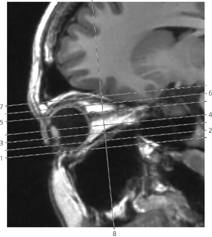
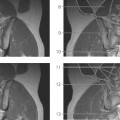
Lines #1–7 indicate position of sections in the following axial MR series. Line #8 indicates the position of a coronal section. Note that bulbar structures do not correspond to the indicated planes in some of the images due to ocular movements during the period of examination.
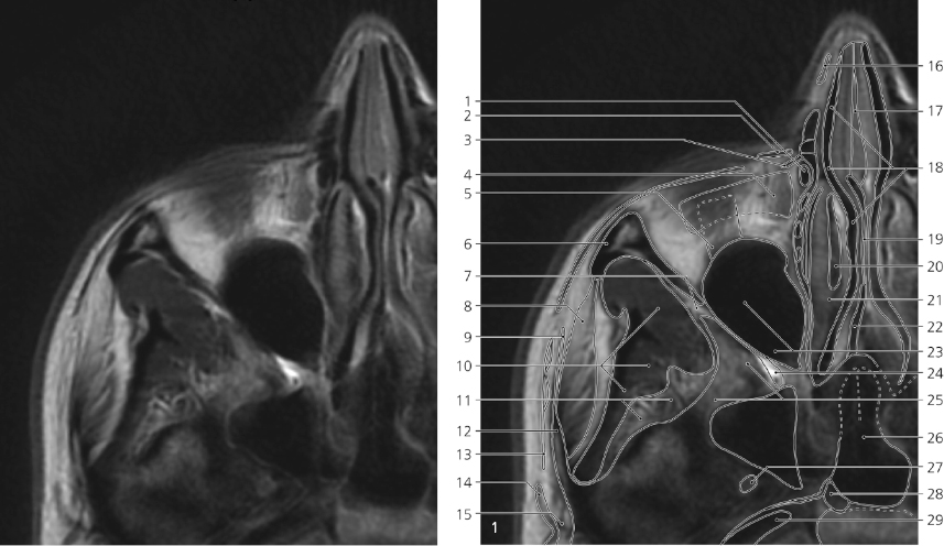
Orbita, axial MR
Lacrimal sac →
Levator labii superioris alaeque nasi
Orbicularis oculi (lacrimal part) →
Inferior oblique
Inferior rectus →
Zygomatic bone →
Inferior orbital fissure →
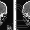
Masseter
Temporal fascia (superficial layer) →
Temporalis →
Pterygoid venous plexus
Zygomatic process of temporal bone
Temporo-parietal fascia →
Anterior auricular muscle →
Superficial temporal artery →
Nasal cartilage →
Nasal septum (cartilaginous part) →
Middle nasal meatus →
Ethmoidal bone (perpendicular plate) →
Middle nasal concha
Mucosa of middle nasal concha
Vomer →
Maxillary sinus →
Pterygopalatine fossa →
Greater wing of sphenoidal bone (with air cell) →
Sphenoidal sinus →
Foramen ovale
Foramen lacerum
Internal carotid artery (in carotid canal) →
Only gold members can continue reading. Log In or Register to continue