Chapter 22 Orthopantomography and cephalometry
Orthopantomography (OPT or OPG) or dental panoramic tomography (DPT)
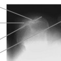
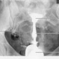
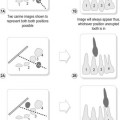
This technique requires the use of a specialised OPT unit (Fig. 22.1A,B), the tomographic principle being that which is used to produce the image of the full mouth and its dentition. The moving tube effectively blurs out the shadow of overlying structures by placing the dental arch in the axis of the tomographic movement. Structures not lying within this axis are effectively blurred, and so their detail does not overlie the image of the teeth and mandible. However, the area of interest does show some element of unsharpness compared to radiographic images of other body parts when a non-moving tube is used.
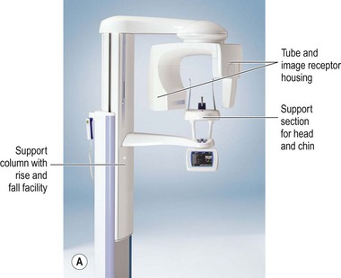
The technique opens out the image of the dental arch to appear in a linear arrangement on the final image. It has long been employed in the dental setting and has largely replaced full mouth periapical examinations. As mentioned in Chapter 18, the OPT examination can be used to demonstrate the temporomandibular joints and mandible.
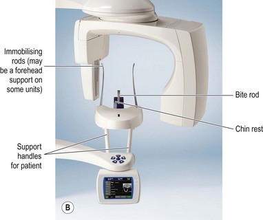
Positioning (Fig. 22.2)
• A disposable bite rod is inserted into the chin rest, or a disposable plastic cover is applied to the permanent bite rod
• The patient is seated or standing with their chin resting on the chin support and in the correct position to facilitate the dental arch being placed in the correct tomographic plane (with the anthropological baseline and alatragal line horizontal and the head far enough forward, often indicated by slit light indicators as designed by the manufacturer)
• The patient bites with their incisors in the groove on the bite rod, to effect separation of teeth on the image
• The MSP is vertical and perpendicular to the bite rod. The height of the unit is adjusted until the occlusal plane is horizontal (assessed by checking that the alatragal line or anthropological baseline are horizontal)
• The patient holds onto the support handles and is asked to step forward slightly to bring the cervical spine vertical. If seated, their chair is pulled forward by the radiographer. Throughout this manoeuvre the head must not tilt or rotate and the chin must not lift or drop
Related posts:

Stay updated, free articles. Join our Telegram channel

Full access? Get Clinical Tree


