Clinical Presentation
The patient is a 46-year-old female with dwarfism who suffers from chronic degenerative joint disease involving the large joints. The patient recently had high cervical myelopathic symptoms including neck pain, bilateral hand numbness associated with neck flexion, and weakness of the upper and lower extremities. She describes having recent episodes of intense tingling sensation that radiates down the spine (positive L’Hermitte’s sign). On physical examination, neck motion is limited in all directions. She has mild distal upper extremity weakness bilaterally and moderate bilateral lower extremity weakness, mainly involving the hip flexors. Plantar flexion ability is diminished. There is a weak positive bilateral Babinski sign. Deep tendon reflexes are normal. No bladder or bowel dysfunction is present.
Imaging Presentation
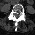
Cervical spine computed tomography (CT) reveals an ossicle superior to a hypoplastic dens consistent with os odontoideum. The anterior arch of C1 is hypertrophic, whereas the posterior arch of C1 is diminutive. Both the anterior and posterior C1 arches contain a midline cleft ( Figs. 52-1 to 52-3 ) . Magnetic resonance (MR) imaging further reveals thickening of the cruciform ligament, which impinges upon the ventral cord surface at the C1-2 level. The anteroposterior (AP) diameter of the central canal is markedly narrowed (5 mm) with the neck in neutral position. Abnormal T2 signal hyperintensity is demonstrated in the spinal cord at the C1-2 level secondary to either cord edema or myelomalacia from cord compression ( Fig. 52-4 ) .


Discussion
Os odontoideum is a term first used by Giacomini in 1886 to describe a potentially unstable condition, whereby the odontoid process is separated from the body of C2. Os odontoideum is an uncommonly encountered condition in routine practice, but when present, is often discovered as an incidental finding on CT or MR scans of the cervical spine. Patients with os odontoideum may or may not have symptoms referable to the upper cervical region. Symptomatic cases of os odontoideum have upper neck pain, stiffness, or neurologic dysfunction secondary to atlantoaxial instability with cord compression.
Embryologically, the odontoid process develops from the fourth occipital sclerotome and the first cervical sclerotome. The C2 vertebral body is formed from the second cervical sclerotome. The normal odontoid process (dens) develops from three ossification centers: a tiny terminal ossification center, the ossiculum terminale, and two larger, columnar-shaped ossification centers that form the majority of the odontoid process including its base. The ossiculum terminale develops from either mesenchyme from the fourth occipital sclerotome or proatlas (or both) and is not ossified at birth. The normal ossiculum terminale appears at age 3 and fuses to the remainder of the dens by age 12. The base of the odontoid process forms the superior portion of the C2 vertebral body centrally. The two basal columnar-shaped ossification centers of the dens begin to fuse in the sixth fetal month. The inferior portion of the dens begins to ossify in the seventh fetal month and ossification progresses from caudal to cephalad. At birth, the cephalad portion of the odontoid is usually not ossified. By 3 to 4 years of age, the odontoid process is completely ossified, and by age 6 it is fused to the C2 vertebral body in most people. Some unossified cartilage may persist into adult life in the center portion of the dens and in the dental synchondrosis (sometimes called the C1-2 disc anlage ), located at the junction of the C2 body and odontoid base.
Although relatively uncommon, os odontoideum is said to be the most common anomaly of the odontoid process ( Figs. 52-1 to 52-5 ) . Other anomalies of the dens also occur. The dens may be developmentally hypoplastic, appearing as a small stump at the superior portion of the C2 body. Rarely, the odontoid process is aplastic. However, true aplasia of the odontoid process would mean that there is complete absence of the dens including its base and therefore some doubt that true odontoid aplasia even exists. In patients with atlanto-occipital assimilation, the atlas is incorporated into or partially fused with the skull base; the dens is usually enlarged and elongated in this condition ( Fig. 52-6 ) . Atlantoaxial instability may be present with any anomaly of the C1-2 articulation.


Stay updated, free articles. Join our Telegram channel

Full access? Get Clinical Tree


