Clinical Presentation
The patient is a 68-year-old woman who presented with instability and tinnitus. She has fallen several times but still ambulates without the use of a walker or cane. She states that she occasionally “loses control of her legs” for no reason, which causes her to fall. She feels that her instability has been occurring for at least 5 years and has progressed over the last year.
Imaging Presentation
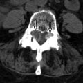
Axial soft tissue and bone algorithm computed tomography (CT) images at the C2 level show bony densities along both the anterior and posterior aspect of the C2 vertebral body. The posterior bony density causes right ventrolateral impression upon the spinal cord. The posterior bony density has corticomedullary continuity with C2 ( Fig. 55-1 ) .

Discussion
Osteochondromas are the most common benign tumors of the long bone. They comprise 8.5% of all osseous tumors and 36% of all benign bone tumors. Osteochondromas are actually considered hamartomas rather than true neoplasms. There are two presentations of osteochondroma: single and sporadic or multiple and hereditary. Multiple exostosis can occur sporadically (25% of cases) but usually results (75% of cases) from a hereditary autosomal dominant disorder known as hereditary multiple exostosis , osteochondromatosis , Bessel-Hagel syndrome, or diaphyseal aclasis . The single and sporadic form affects the spine in 3% of cases, and the multiple form in 7% to 12% of cases. Most spinal lesions are the solitary type, with spinal osteochondromas comprising 0.4% of all spinal tumors. Approximately 10% to 15% of osteochondromas develop secondary to radiation exposure with an average latency of 8 years. Osteochondromas are common in children who received radiation doses of greater than 25 Gy when they were younger than 2 years of age. Osteochondroma is the only bone tumor that can be reproduced experimentally. Solitary osteochondromas affect male patients more than females with a ratio of 2.5 : 1 and an average age at presentation of 30 years. Patients with multiple exostoses present at an average age of 20 years.
Approximately 50% of spinal osteochondromas occur in the cervical spine. The most common level affected is C2, followed by C3 and C6. It has been postulated that microtrauma to the cervical vertebra resulting from its greater mobility might explain the higher incidence of osteochondroma in the cervical region. The thoracic spine is the next most common site, typically affecting T8 followed by T4. Spinal lesions most frequently occur in the posterior arch, usually arising from the tip of a spinous process or transverse process.
Approximately 30% of solitary spinal osteochondromas cause spinal cord compression as opposed to 50% in the multiple form. Clinical symptoms are often slow and progressive because of the slow nature of the growing neoplasm. However, some patients can have an acute presentation of symptoms after a sudden hyperextension or fall in the face of an already compromised spinal canal. Although rare, patients with a cervical osteochondroma can die suddenly from spinal cord compression. Patients can exhibit symptoms of radicular pain, claudication, and/or myelopathy.
Malignant transformation of an osteochondroma to a chondrosarcoma is a well-known complication that is seen in 1% to 5% of patients with solitary osteochondromas and in 10% to 25% of those with the multiple hereditary form. Malignant transformation should be suspected if the cartilaginous cap is greater than 2 cm in thickness, if the lesion recurs after resection or grows after skeletal maturity, or if there is new onset of pain or a sudden increase in lesion size.
Imaging Features
A mature osteochondroma consists of a stalk and body composed of mature bone, which is covered by a cartilaginous cap that never ossifies and is never seen on plain radiography. Corticomedullary continuity of the osteochondroma with the parent bone is pathognomonic. On plain films, an osteochondroma appears as a pedunculated or sessile bony projection. At the point of attachment, the cortex of the bone or origin flares into the cortex of the osteochondroma ( Fig. 55-2 ) . This is considered a pathognomonic finding. Due to multiple overlapping structures in the spine, plain films are often insufficient for diagnosis. Computerized tomography (CT) readily demonstrates the corticomedullary continuity of the osteochondroma with the parent bone where the cortex can be followed as a single line and the medullary cavities of the parent bone and osteochondroma are as one ( Fig. 55-3 ) . Flocculent calcification or an “arcs and rings” matrix may be seen in the osteochondroma. Although CT can demonstrate the osseous component of an osteochondroma, it fails to demonstrate the cartilaginous cap in the majority of cases, and when it does, it often underestimates the size of the cartilaginous cap. This makes it difficult to distinguish between an osteochondroma with a thick cartilaginous cap and a chondrosarcoma with a thin cartilaginous cap.


