Osteosarcoma, Chordoma, and Chondrosarcoma


Osteosarcoma, Chordoma, and Chondrosarcoma


Shiao Y. Woo

Edward C. Halperin


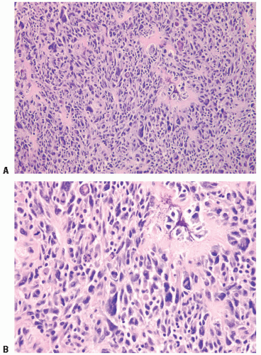
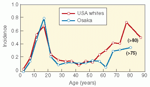
Osteosarcoma is the most common primary malignant bone tumor in children. The tumor derives from boneforming mesenchyme (1, 2, 3, 4, 5, 6, 7, 8, 9) (Fig. 10-1A,B). The majority of cases occur in the second decade of life, and there is a male predominance. The incidence is one to three new cases per million per year (Fig. 10-2) (10).

Inactivation of the retinoblastoma (Rb) gene may be important for osteosarcoma formation. Rb is a tumor suppressor gene that is discussed in detail in Chapter 5. A common karyotype change in osteosarcoma is deletion of the short arm of chromosome 17, where the p53 gene is localized; p53 is a nuclear phosphoprotein with properties of a tumor suppressor gene (11). Genetic studies on human, canine, and murine osteosarcomas have revealed mutation of c-kit (12) and TP53 (13) genes, over-expression of urokinase plasminogen activator/uPA receptor (14), CRM1 (15), Ezrin (16), alpha V integrin and vascular endothelial growth factor (17), runt-related transcription factor Runx2 (18) and URG4 gene(associated with cell proliferation) (19), increased phosphorylation of cJun, JNK and ERK1/2 (20), and expression of cyclooxygenase (COX-2) (21) and somatostatin receptors (22). Some of these genetic alterations may have oncogenic or prognostic significance and may also be potential therapeutic targets. Highresolution array comparative genomic hybridization in combination with interphase fluorescence in situ hybridization has been shown to detect chromosomal instability and genomic imbalance that have been correlated to response to chemotherapy (24).


Figure 10.1 A,B: Histopathology of osteosarcoma: low power (A) and high power (B).


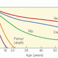
Figure 10.2 Osteosarcoma incidence per 100,000 population per year by age. The peak for older people is associated with Paget disease. (Reproduced from Ishikawa Y, Tsukuma, H, Miller RW, et al. Low rates of Paget’s disease of bone and osteosarcoma in elderly Japanese. Lancet. 1996;347:1559, with permission.)

Second malignant neoplasms can occur in patients with osteosarcoma treated with surgery alone or with surgery and chemotherapy, perhaps as a result of these genetic abnormalities (24, 25, 26). Osteosarcoma may develop in long-term survivors of heritable Rb. These secondary osteosarcomas may arise in or outside the irradiated field and in children treated without radiotherapy (see Chapters 5 and 20).


SIGNS, SYMPTOMS, EVALUATION, AND STAGING

Osteosarcoma usually occurs in the metaphyses of the long bones, especially around the knee joint. The bones most commonly involved are the femur (approximately 40% of cases),
tibia (15%), and humerus (15%) (Fig. 10-3) (1,3,4,10,27). The usual clinical presentation is swelling or pain. A few patients present with a pathologic fracture.


Figure 10.3 Skeletal distribution of primary osteosarcomas in patients treated on the Neoadjuvant Cooperative Osteosarcoma Study Group protocols of the Cooperative German-Austrian-Swiss Osteosarcoma Study Group. (Modified from Bielack S, Kempf-Bielack B, Delling G, et al. Prognostic factors in highgrade osteosarcoma of the extremities or trunk: an analysis of 1702 patients treated on Neoadjuvant Cooperative Osteosarcoma Study Group protocols. J Clin Oncol. 2002;20: 776-790, with permission.)

The tumor has a typical appearance on conventional radiographs. There are poorly defined margins, interrupted periosteal new bone, and soft tissue invasion. Where the bony cortex is penetrated at the edge of a tumor, there may be a periosteal elevation and vertical spicule formation (Codman’s triangle) (3,4,27). Computed tomography (CT) and magnetic resonance imaging (MRI) help delineate the intramedullary extent of tumor as it tracks along the marrow cavity and the soft tissue extent of tumor (4,28) (Fig. 10-4). The bone scan has nearly 100% sensitivity for the presence of malignant bone tumor, although the specificity is less. On the bone scan one may observe osteoblastic activity in the shaft of the long bone proximal to the primary tumor. This may represent reactive change and not indicate the presence of malignancy (4). Thallium scintigraphy also aids in tumor localization. A surgeon may order an angiogram to determine tumor vascularity, detect vascular displacement, determine the relationship between vessels and the tumor, and identify vascular anomalies. The most common sites of distant metastases are the lungs and bones (3,6,27, 28, 29, 30, 31). To evaluate pulmonary metastases, a plain chest radiograph is used to identify chest nodules or cannonball lesions. A CT of the chest can be used to assess the presence or absence of small nodules (31).

If we are to improve the treatment of osteosarcoma, it will be important to adapt therapy to the individual patient’s prognostic factors (32). The conventional means of doing this is a staging system. The presence or absence of metastases and the use of histologic subtyping do help predict prognosis (Table 10.1). A variety of factors have been investigated as potential prognostic factors. As will be discussed later in this chapter, histologic response to neoadjuvant chemotherapy is useful in predicting outcome. However, information of this type is available only after chemotherapy and surgery. Clinical features with predictive value in determining outcome include the duration of presenting symptoms (shorter is worse), tumor size (larger is worse), location of the primary tumor (head, spine, rib, and pelvic sites are worse), and weight loss of >10 lb. Tumor size seems strongly predictive of outcome and may be calculated as either absolute tumor length (>10 cm), relative tumor length given as the proportion of tumor length to the overall length of the involved bone (more than one third of the involved bone), or absolute tumor volume (>70 or 150 cm3) (35,36).


Figure 10.4 An 18-year-old right-handed high school senior presented to medical attention when his tennis game and weight-lifting activities were inhibited by pain in his left shoulder. Diagnostic images showed a mixed sclerotic and lytic mass in the proximal left humerus. Incisional biopsy made the diagnosis of osteosarcoma. The patient was treated with induction chemotherapy in the hope of producing a substantial tumor response eventually leading to limb-sparing surgery. Unfortunately, within 2 years of the initial diagnosis widespread pulmonary metastases developed, refractory to chemotherapy, which led to the patient’s death.

There is no widely used staging system for osteosarcoma. Some clinicians simply separate patients into two groups: those with localized disease and those with metastatic disease. The system developed by Enneking and adopted by the Musculoskeletal Tumor Society is used by some orthopedic oncologists. However, there are few low-grade osteosarcomas, so in this system most tumors are stage II or III (Table 10.2). One study from the European Osteosarcoma Intergroup suggested that patients with chondroblastic tumors had a slightly superior survival to those with other histologies, but this difference did not reach statistical significance and awaits confirmation in other research (34).

The Cooperative German-Austrian-Swiss Osteosarcoma Study Group (COSS) has provided an extensive evaluation of prognostic factors in high-grade osteosarcoma of the extremities or trunk. In an evaluation of 1702 patients treated on
neoadjuvant osteosarcoma protocols, the investigators found that the long-term survival of patients with limb primaries was superior to that of those with axial primaries, patients who presented without metastases had higher survival rates than those who presented with metastases, patients with extremity sarcomas occupying less than one third of the bone had higher survival rates than those with sarcomas occupying more than one third, and distal extremity tumors had a superior outcome to proximal tumors. The response to chemotherapy (i.e., the amount of tumor necrosis at the time of resection in a setting of neoadjuvant chemotherapy) also was a highly important predictor of outcome along with the extent of surgical resection (Table 10.3) (37, 38, 39).


Table 10.1 A System for Osteosarcoma Subclassification by Histology and Origin


Centrally Located Tumor


Primary tumors


Conventional: About 75% of patients fall in this category. This group may be additionally subdivided based on the predominant matrix pattern into the following three subgroups:


Osteoblast, 14.4 (54%)


Chondroblastic, 9.6 (70%)


Fibroblastic (spindle cell stroma with a herringbone pattern similar to that seen in fibrosarcoma), 10.7 (57%)


Telangiectatic (characterized by a purely lytic radiographic appearance and a macroscopic and microscopic resemblance to aneurysmal bone cyst), 14.5


Small cell


Malignant fibrous histiocytoma subtype


Low-grade intraosseous (a rare low-grade fibro-osseous lesion often confused with fibrous dysplasia)


Multicentric


Gnathic


Secondary tumors


Associated with Paget disease, 64.2


Radiation-induced


Associated with other benign pre-existing condition (i.e., fibrous dysplasia)


Juxtacortical tumor, 1.0


Parosteal (often arises from the posterior distal femur in older patients. Generally it is a low-grade spindle cell tumor with well-formed, parallel trabecular bone. Some patients may dedifferentiate into a higher grade lesion)


Periosteal (typically involves femur or tibia)


High-grade surface


The death rates per 100 patient-years, correlated with histologic group and calculated by Taylor et al. (33), are shown in bold type, and the 5-year survival rates from the European Osteosarcoma Intergroup (34) are in parenthesis. The death rates for the three subtypes of conventional osteosarcoma are similar. There is some controversy in the literature concerning the prognostic importance of the telangiectatic variant. Most authorities now believe that with aggressive therapy, there is no prognostic difference attached to this diagnosis variant. It is generally agreed that the prognosis for patients with juxtacortical tumors is better and that the prognosis for tumor associated with Paget disease, an entity seen in adults, is decidedly worse. From Refs. 1,4,7,9,33,35, and 43, with permission.


Table 10.2 The Enneking Staging System for Osteosarcoma


Stage I


Low Grade


IA. Confined to bone of origin


IB. Extension beyond bone of origin


Stage II


High Grade


IIA. Confined to bone of origin


IIB. Extension beyond bone of origin


Stage III


Metastatic Disease


IIIA. Confined to bone of origin


IIIB. Extension beyond bone of origin


Modified from Damaron JA, Pritchard DJ. Current combined treatment of high-grade osteosarcomas. Oncology 1995;9:327-350; and Ennedking WF, Spanier SS, Goodman MA. A system for the surgical staging of musculoskeletal sarcoma. Clin Orthop. 1980;153:106-120, with permission.


SELECTION OF THERAPY


Surgery

The major modalities of therapy are surgery, radiation therapy and chemotherapy.


Local Disease

The biopsy site should be selected to allow access to the infiltrating edge of the tumor. In general, either minimal or no cortical bone should be removed in order to reduce the risk of pathologic fracture (42, 43, 44, 45, 46).

The classic definitive operative procedure is an amputation above the region of the affected bone or a disarticulation at the joint above the lesion. Traditional teaching is that if one resects the bone beyond the site defined by all radiographs as the most proximal extent of disease and if the margins are pathologically negative, the chance of stump recurrence is negligible (44,45). In recent years surgical treatment for local osteosarcoma has changed. New limb salvage procedures have been performed with gratifying results (47).


Table 10.3 Influence of the Histologic Response to Neoadjuvant Chemotherapy on Survival in Osteosarcoma


Group (Reference)


Response


n


Survival


p


Istituto Ortopedico


≥ 90% necrosis


621


76% 5-year OS


0.0001


Rizzoli (38)


< 90% necrosis


437


48% 5-year OS


Pediatric Oncology


≥ 90% necrosis


26


73% 5-year EFS


0.027


Group 8651 (41)


< 90% necrosis


16


44% 5-year EFS


EFS, event-free survival; OS, overall survival.


Limb-sparing operations may be selected if there is no evidence of neurologic or vascular compromise by the local tumor, if the surgeon believes that he or she can obtain an adequate margin around the primary osteosarcoma, and if there is a plan for reconstruction that will provide better function than amputation. Relative contraindications to limb sparing include the presence of a pathologic fracture, a poor response to neoadjuvant chemotherapy, or skeletal immaturity that will lead to significant limb growth discrepancies (48).

What is an adequate surgical margin for local management of osteosarcoma? A radical margin entails removal of the entire bone of origin with accompanying soft tissue involvement, as is achieved by a hip disarticulation for a distal femoral tumor. A wide margin is defined as excision of the tumor with a cuff of surrounding normal tissue, and a marginal margin entails excision of the tumor and its surrounding reactive pseudocapsule. Local control is improved by the adequacy of the surgical margin and the response to neoadjuvant chemotherapy (Table 10.3). It is generally accepted that limb-sparing surgery has a slightly greater risk of local tumor recurrence than does amputation. Amputation has a slightly greater risk of local recurrence than does disarticulation (Table 10.4). A good chemotherapeutic response may allow the surgeon to have a tighter margin on the primary tumor and exclude more normal tissue from the resection. However, to know before surgery whether a marginal excision is reasonable, one must have an idea of what tumor response was achieved by chemotherapy. Unfortunately, no single imaging study can reliably provide this information, although plain radiographs, CT, bone scan, MRI, and FDG-PET scan can be used to make a reasonable prediction (48,50).


Table 10.4 Local Recurrence Rates for Osteosarcoma as a Function of the Extent of Surgery


Site (Reference)


Type of Operation


Local Recurrence rate (%)


Distal femur (37)


Limb sparing


Above-the-knee amputation


11


8


Hip disarticulation


0


Pelvis (49)


Radical or wide


48


Marginal


70


Intralesionala


92


a Some patients were irradiated.


The reconstruction technique elected after limb-sparing procedures depends on the location of the osteosarcoma, whether there is joint involvement, the extent of bone and soft tissue resection, the patient’s age, the functional demands of the patient and the family, and the prospects for rehabilitation. When the excision is intra-articular (within or involving a joint), reconstruction options include custom segmental total joint replacement, whole segment osteoarticular allograft (i.e., from a cadaveric donor), allograft-prosthetic composite reconstruction, arthrodesis (surgical fixation of a joint; artificial ankylosis) with autologous or allogeneic bone, or arthrodesis with a porous prosthesis allowing host bone ingrowth and bone graft. If a joint is not involved, one can reconstruct with autologous or allogeneic bone, a prosthesis, or a segmental prosthetic spacer. Rotation plasty is also an option in certain situations (2,51, 52, 53).

It is gratifying that, in modern pediatric oncology practice, the child with osteosarcoma may be offered limb-sparing options for local treatment beyond the traditional amputation or hip disarticulation. The previous discussion has cited indications and relative contraindications to limb-sparing surgery, the risk of local recurrence after various forms of surgery, options for reconstruction after tumor resection, and the need for adequate surgical margins. Although limb function after limb-sparing surgery is generally good in many patients, limb-sparing surgery is not for everyone faced with osteosarcoma. Case selection by a skilled orthopedic oncologist, invocation of sound oncologic principles, and adequate rehabilitative services postoperatively are all necessary to achieve the best possible function and cancer control.


Metastatic Disease

Surgery also plays a role in treating osteosarcoma metastatic to the lung. Some patients present with a primary tumor along with limited pulmonary involvement. Aggressive multiagent chemotherapy, surgical management of the primary tumor, and thoracotomy for resection of pulmonary metastases appear to have significantly increased survival in cases that seemed hopeless (Table 10.5) (54). The most common sites of metastases in the relapse of initially localized osteosarcoma are lung and bone (Fig. 10-5). When tumor relapses in the lung, surgical resection of pulmonary nodules may result in a prolonged disease-free interval and, together with aggressive chemotherapy, a small potential for cure (6,25,30,29,44,55,56). In some patients, repeated thoracotomies appear to have prolonged survival in the face of multiple episodes of pulmonary metastases (43). By diagnosing pulmonary recurrences earlier and with limited tumor bulk, thoracic CT may either
open the possibility for a beneficial effect of surgery or simply create a lead-time bias (54).


Table 10.5 Factors Predicting Event-Free Survival in Patients with Primary Metastatic Osteosarcoma in the German-Swiss-Austrian Cooperative Osteosarcoma Study Group Clinical Trials


Factor


n


Five-Year Event-Free Surviva


p


Primary site


Extremity


181


20%


< 0.001


Trunk


21


5%


Number of metastatic organ systems


1


160


21%


< 0.006


> 1


42


10%


Number of metastases


1


38


55%


< 0.001


2-5


69


19%


> 5


91


2%


Isolated lung metastases


Unilateral


46


45%


< 0.001


Bilateral


70


4%


1


24


58%


< 0.001


2-5


41


23%


> 5


57


3%


From Kager L, Zoubek A, Potschger U, et al. Cooperative German-Austrian-Swiss Osteosarcoma Study Group. Primary metastatic osteosarcoma: presentation and outcome of patients treated on neoadjuvant Cooperative Osteosarcoma Study Group protocols. J Clin Oncol 2003;21(10):2011-2018, with permission.


Several variables must be considered when one uses metastasectomy for pulmonary metastases. These include the aggressiveness of the proposed operation, whether they are unilateral or bilateral, the interval between the development of the metastases and the treatment of the primary, the extent of vascular invasion, the presence or absence of hilar lymph node involvement, and whether additional salvage chemotherapy is available after surgery (48,49,57). The role of the bony metastasectomy is not well defined, undoubtedly because the occurrence of bony metastases in the setting of potentially salvageable osteosarcoma is far less common than that of pulmonary metastases (48).

Bielack et al. reviewed the outcomes of 249 consecutive patients with second and subsequent recurrences of osteosarcoma (58). With mainly chemotherapy and surgery, the 5-year overall and event-free survival rates were 32% and 18% for second, 26% and 0% for third, 28% and 13% for fourth, and 53% and 0% for fifth recurrences, respectively.


Radiation Therapy


Prebiopsy

Sweetnam (44) administered low-dose irradiation before the initial biopsy (approximately 10 Gy) to 29 patients in the hope of reducing the viability of cells that might be disseminated into the bloodstream by the biopsy. The 20% overall survival rate, no different from that of historic controls, discouraged additional investigation. Only 2 of 19 patients survived when treated with amputation without prior biopsy.


Primary and Preoperative Treatment


The Cade Technique

In the era before adjuvant chemotherapy, physicians were distressed by the practice of treating the primary lesion with amputation or disarticulation, only to have the young patient die within 6 months of pulmonary metastases. Because the survival rate with surgical ablation alone was only 20%, many limbs were sacrificed in vain. Surgeons and radiotherapists reasoned that if high-dose local irradiation could obtain at least temporary control of the primary tumor, a time interval would be obtained that would allow the selection of cases suitable for a radical surgery. Patients who develop pulmonary metastasis in a 4-6 month waiting period after irradiation would be spared an unnecessary amputation. Those who did not develop pulmonary metastasis after the waiting period would undergo extirpation of the primary tumor. This philosophy was promulgated by English physician Sir Stanford Cade and is called the Cade technique. The 5-year survival rates of 15-20% were equivalent to those achieved in the preadjuvant therapy era with immediate surgical ablation. The delay in surgery appears to have cost no lives (10,56,59, 60, 61, 62, 63, 64, 65, 66, 67). There generally was a reduction in pain and swelling at the tumor site after the first 20 Gy. This response tended to continue for several weeks after completion of radiotherapy. In patients who underwent limb ablation after radiotherapy, a histologic analysis could be performed to assess the presence or absence of viable tumor. The majority of patients had a good or excellent local response (Table 10.6). There were some long-term survivors after aggressive treatment with radiation therapy alone (6,66).


Modern Series of Primary Photon Radiation Therapy

In modern radiotherapy practice, it is rare to be asked to use radiotherapy as the primary local treatment for osteosarcoma except for lesions in inaccessible sites. However, the data acquired with the Cade technique make it reasonable to consider the use of radiation in certain situations. Preoperative radiotherapy has been given in the context of a research
protocol to reduce tumor viability before surgery, increase the probability of performing limb-sparing surgery instead of amputation, or reduce the risk of local recurrence (56,67). In patients with nonresectable primary tumors, such as difficult pelvic bone sites, vertebral column, frontal bones, or base of skull, and in patients who refuse definitive surgery, consideration should be given to precision high-dose irradiation. Modern photon techniques use three-dimensional computerized treatment planning and intensity-modulated radiation therapy. Neutron or proton beams can improve local control in certain circumstances and are discussed later in this chapter (71). There is also precedent for high-dose preoperative irradiation and rapid surgery, preoperative radiotherapy with local hyperthermic perfusion (72), intraoperative electron beam therapy (73), and radiotherapy with intra-arterial infusion of a radiosensitizer (74).


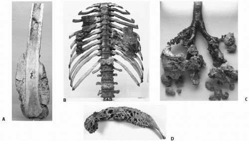
Figure 10.5 John Hunter (1728-1793) was an extraordinary surgeon, anatomist, teacher, and collector. His magnificent collection of scientific specimens was purchased by Parliament and placed in the custody of the Company of Surgeons, renamed the Royal College of Surgeons in 1800. These illustrations of osteosarcoma, taken from the Hunterian Museum, Lincoln’s Inn Fields, London, appear by permission of the president and council of the Royal College of Surgeons of England. In November 1786, Hunter encountered a patient with “a hard swelling of the lower part of the thigh, as it were beginning from the knee … the part began evidently to enlarge … and was attended with more pain as it enlarged … and the pain was now exhausting him much.” An amputation was done. Hunter described an “osteoid sarcoma” of the distal femur (A) and also noted the intramedullary spread of the tumor: “A short distance below the site of amputation there is a second hemispherical tumour in the medullary canal … above the main growth, the disease having extended within the canal, in this case, for an unusual distance beyond the limits of the external swelling.” Four weeks after the amputation the patient “began to complain of a difficulty in breathing, but not attended with the least pain … he began to lose his flesh and sink gradually, his breathing being more and more difficult … he died; living only 7 weeks after the operation.” On autopsy, “bony tumors were found in the cellular membrane of the lungs, upon the pericardium, and some very large ones of the pleura, adhering to the ribs, and upon the anterior surface of the vertebrae of the back” (B,C). Hunter noted that “when the leg was amputated he had not the least symptom of any disease in the chest” but deduced that the lung metastases “had taken place a considerable time before the symptoms took place.” As for the osteoid appearance of the tumor, Hunter wrote that “one can figure to themselves a reason why the tumour which formed on the outer surface of the thigh-bone might become bony, because it might acquire that disposition from the bone it surrounded; but, from these tumours formed in the chest becoming bone, shows it was the nature of the tumours themselves.” The remaining figure (D) is “the osseous part of an osteosarcomatous tumour” from “the rib of a Horse” from Hunter’s collection. (Descriptions of this material are found in Descriptive catalog of the pathological series in the Hunterian Museum of the Royal College of Surgeons of England. Edinburgh and London: E. & S. Livingstone, 1972; Part I:133-138; Part II:75-77. Biographical material is from Allen E. Hunterian Museum [pamphlet], Royal College of Surgeons, 1974.)

Data on photon irradiation as primary treatment for osteosarcoma, in lieu of surgery and in conjunction with aggressive chemotherapy, are available from Albrecht et al. from Berlin (59). They described seven patients with osteosarcoma who were treated from 1977 to 1992 according to contemporary COSS protocols (Table 10.7). Six patients refused the appropriate amputation or rotation plasty. One had a primary tumor described as inoperable. Each patient received 50-70 Gy of conventionally fractionated photon irradiation. The patient with the inoperable tumor died within 1 year of initiation of treatment. The remaining six patients are alive without evidence of recurrent disease 2-18 years after treatment (mean follow-up, 11 years). Three of the six survivors ultimately suffered a pathologic fracture 8-12 months after 50-70 Gy, and one of these subsequently had an amputation. This small clinical series suggests that photon radiotherapy in
conjunction with chemotherapy may be used to manage osteosarcoma if appropriate surgery is impossible or refused (59). Whether such patients are best treated with photons, neutrons, or protons is a matter for debate.


Table 10.6 The Cade Technique: Management of Osteosarcoma with Primary Irradiation or Primary Irradiation and Selected Delayed Amputation (Excluding Parosteal Tumors)


Authors Listed in Order of Increasing Dosage (References)


Dosage (Gy)


Clinical or Pathologic Local Response


Survival


Jenkin et al. (64,65)


50-60


Complete, 11%


Partial, 63%


None, 26%


2-year: 16%


van den Brenk et al. (81)


60-70, then 40-50 every 6 months


Severe limb deformity in long-term survivors


5-year: 12% (includes cases of chondrosarcoma and fibrosarcoma)


Lee et al. (56,66)


70-80


33% totally destroyed


33% doubtful viability


33% capable of growth and perhaps dissemination


5-year: 22%


Poppe et al. (10)


70-80


70% considerable radiation effect


19% moderate effect


5-year: 18%


Gaitan-Yanguas (63)


10-100


11% slight or no effect


44% no residual tumor


Allen and Stevens (60)


79-100


56% residual tumor


60% reclassification


86% tumor sterilization


60% NED at 30-114 mo


Caceres and Zaharia (61)


80-120


16% remaining viable tumor


56% severe radiation damage


28% total tumor destruction


35% no tumor destruction


5-year: 18% (no difference from historical surgical controls, 22%)


Phillips and Sheline (67)


50-120


65% tumor in specimen


NED, no evidence of disease.


Some patients with unresectable osteosarcomas are treated with conventional external beam irradiation. The COSS described a series of patients with osteosarcoma of the spine who were treated with intralesional or marginal resections and also received photon irradiation, neutron beam treatment, or samarium. In the analysis of patients who underwent either incomplete surgery or no surgery, 7 received postoperative irradiation and 10 did not. The seven patients who received irradiation had a slightly higher long-term survival (50%) than those who did not (10%, p = 0.059) (84).

In 2003, COSS investigators described a group of patients with pelvic osteosarcomas treated between 1979 and 1998. Of 30 patients with intralesional surgery or no primary surgery, 11 received radiotherapy (65-68 Gy with photons, 2 were treated with neutrons, and 1 also received 153Sm). The 5-year overall survival of the irradiated patients (16%) was superior to that of those not irradiated (0%, p = 0.0033) (85).

Machak et al. (86) reported a retrospective review of 31 patients with limb osteosarcoma who refused definitive surgery and were treated with cis-platinum, doxorubicin (IV or IA), or both. All patients received 40-68 Gy at 2.5-3 Gy per fraction, one fraction per day, or 1.25-1.5 Gy twice daily. With a median follow-up of 39 months, the predicted 5-year overall survival was 61%. Local progression-free survival was 56% (40% for 50 Gy or less, 77% for more than 50 Gy). There were five fractures, one skin necrosis, and one osteomyelitis. It is conceivable that use of neoadjuvant radiotherapy with chemotherapy increased the proportion of good responders of the time of surgery (87).

Hirano et al. (52) from Nagasaki University sandwiched 30 Gy (2-3 Gy per fraction) in the midst of preoperative chemotherapy in 15 patients with osteosarcoma. Histology of the resected specimens showed a tumoricidal effect in nine patients and a lesser effect in three.

Wagner et al. used a combination of short-course preoperative irradiation (19.8 Gy), surgical resection, intraoperative 90Y plaque brachytherapy to at-risk areas of the dura when applicable, and postoperative irradiation with sequential cone-downs to 50.4-58.2 Gy (total 70.2-77.4 Gy) to treat bone tumors mainly involving the spine and pelvis (68). For patients with osteosarcoma, the 5-year local control, disease-free survival and overall survival were, respectively, 75%, 50%, and 67%.


Table 10.7 Significant Recent Cooperative Group Trials Addressing the Management of Localized Osteosarcoma


Study Name and Number of Eligible Patients


Primary Objective


Treatment Arms


Results


Comments (Reference)


Pediatric Oncology Group (POG)


POG-8651 (n = 100)


To determine whether chemotherapy administered before and after definitive surgery is superior to surgery followed by adjuvant chemotherapy


Arm A: HDMTX + AP, then surgery, then HDMTX + BCD + ADR + AP


Arm B: surgery, then HDMTX + AP + BCD


The 5-year survival is 76% for patients assigned to neoadjuvant chemotherapy and 79% for patients treated with the more traditional approach (p = 0.6)


There is no evidence of an advantage in event-free survival or survival for either treatment arm (41,78)


European Osteosarcoma Intergroup (EOI, a combination of the European Organization for Research and Treatment of Cancer and the Medical Research Council)


EOI-80831 (n = 307 registered, 207 evaluable, 163 completed allotted chemotherapy)


This study began as a randomized phase II toxicity and response trial of two short, intensive chemotherapy programs


Surgery, randomization of chemotherapy of AP vs. AP + MTX


6-year survival: AP 65% + MTX 50% (p = 0.10)


6-year disease-free survival: AP 58% vs. AP + MTX 40% (p = 0.02)


The dosage intensity of AP was greater in the two-drug arm than the three-drug arm. This may have produced the superior outcome. Disease-free survival was better for patients planned for conservation surgery than amputation (76)


EOI-80861 (n = 407, 391 eligible)


To compare short, intensive chemotherapy with complex, longer-duration program based on the Rosen T10 program


Arm A: AP three cycles, then surgery, then three more cycles Arm B: ADR + V + HDMTX, then surgery, then BCD + ADR + V + HDMTX + AP


The median follow-up is 5.6 years. Survival in both groups is almost identical. 5-year progression-free survival is 44%


AP appears to be as effective as the more complicated program at a lower cost (75,77)


Scandinavian Sarcoma Group (SSG)


SSG-VIII


To increase the number of good responders to neoadjuvant therapy with intensified


HDMTX + AP, then surgery; good responders get HDMTX + AP postoperatively, poor


The median follow-up is 6.9 years. The year event-free survival is 61%


Female sex, small tumor volume, and high serum MTX levels predict better


Children’s Cancer Group (CCG)


CCG-741 (n = 166)


To compare HDMTX with moderate-dose MTX in the context of a multiagent chemotherapy program


Surgery, ADR, then randomize to arm: Arm A, HDMTX + AV; Arm B, moderate-dose MTX + AV


38% disease-free survival at 48 mo. No difference between the two arms (p > 0.5)


Lower disease-free survival was associated with the presence of spontaneous tumor necrosis at presentation (78)


CCG-782 (n = 232)


To use histologic response of the primary tumor to neoadjuvant chemotherapy to determine postoperative chemotherapy


Biopsy, then HDMTX + AV + BCD, then surgery. All patients without local progression received BCD + AV + HDMTX. Then, if< 95% necrosis, receive BCD + HDMTX + AV


5-year event-free survival was 53%. Patients with local disease progression in the induction phase had 48% 3-year event-free survival


A poor prognosis is associated with an evaluated alkaline phosphatase at diagnosis or a primary tumor in the proximal humerus or proximal femur (40,79,76)


Cooperative Osteosarcoma Study (COSS) Group of the German Society of Pediatric Oncology


COSS-77 (n = 68)


Improved survival from adjuvant chemotherapy


Surgery, then HDMTX + AV


46% 14-year metastasis-free survival


Event-free survival was 48% for good responders and 48% for poor responders (80,81)


COSS-80 (n = 101)


Improved survival with neoadjuvant chemotherapy


Arm A: ADR + HDMTX + P, then surgery, then ADR + HDMTX + P


Arm B: ADR + HDMTX + BCD ± β-IFN


5-year event-free survival: Arm A = 66%, Arm B = 64%


There was no difference in outcome between the two chemotherapy arms. There was no difference associated with the use of β-IFN (40,79, 80, 81)


COSS-86 (n = 153)


Stratified by risk group. High risk was large tumor, chondroid matrix, or poor bone scan response at week 5


Low risk: ADR + HDMTX + P, then surgery, then ADR + HDMTX + P


High-risk Arm A: ADR + HDMTX + intra-arterial IP, then surgery, then ADR + HDMTX + IP


High-risk Arm B: Same as Arm A except IP is intravenous


77% 5-year metastasis-free survival, 66% 10-year metastasis-free survival, 72% 10-year overall survival


Response rate is > 76%, better than in previous COSS studies (82)


Children’s Oncology Group (COG)


CCG-7921, POG-9351 (n = 662)


To compare three-drug chemo with four-drug chemo. To determine if addition of MTP improves EFS and OS


Reg A: AP + MTX ± MTP


Reg B: AP + MTX + I ± MTP


Similar EFS and OS between Reg A and B without MTP


MTP-improved 6-year OS from 70% to 78% (p = 0.03)


Addition of ifosfamide was of no benefit. Addition of MTP improved survival


ADR, doxorubicin; AP, doxorubicin and cis-platinum; AV, doxorubicin and vincristine; BCD, bleomycin, cyclophosphamide, and actinomycin D; β-IFN, beta-interferon; EI, etoposide and ifosfamide; HDMTX, high-dose methotrexate; IP, ifosfamide and cis-platinum; MTX, methotrexate; P, cis-platinum; V, vincristine: MTP, muramyl tripeptide.

Only gold members can continue reading. Log In or Register to continue

Stay updated, free articles. Join our Telegram channel

Jun 19, 2016 | Posted by in GENERAL RADIOLOGY | Comments Off on Osteosarcoma, Chordoma, and Chondrosarcoma

Full access? Get Clinical Tree

Get Clinical Tree app for offline access