Fig. 26.1
(a, b) T2-weighted coronal oblique MRI images demonstrate tearing of the RUHL and LCL complex off the posterior aspect of the lateral epicondyle of the humerus, laxity in the annular ligament around the radial head, and edema in the bone of the lateral epicondyle and surrounding musculature. a also shows tearing in the mid-substance of the MUCL with avulsion distally off the sublime tubercle of the ulna (a, b: Published with kind permission. Copyright © Felix H. Savoie, III, MD)
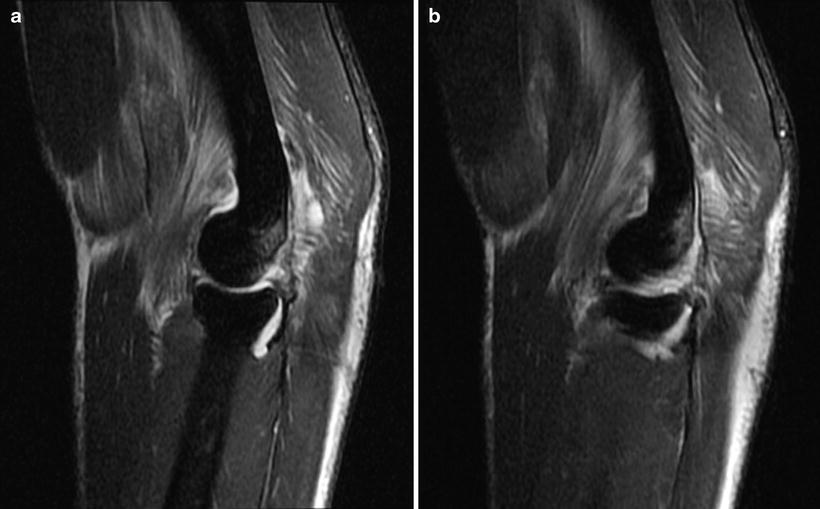
Fig. 26.2
(a, b) T2-weighted sagittal oblique MRI images demonstrate tearing of the RUHL and LCL complex off the posterior aspect of the lateral epicondyle of the humerus, laxity in the annular ligament, edema in the bone posteriorly, and edema in the brachialis muscle anteriorly and triceps muscle posteriorly (a, b: Published with kind permission. Copyright © Felix H. Savoie, III, MD)
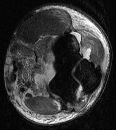
Fig. 26.3
A T2-weighted axial MRI image shows tearing of the RUHL off the posterior aspect of the lateral epicondyle in the upper right corner of the image. The left side of the image demonstrates tearing in the anterior capsule, edema in the brachialis, and hematoma in the anterior compartment of the elbow (Published with kind permission. Copyright © Felix H. Savoie, III, MD)
MRI revealed concentric reduction of the ulnohumeral joint with no interposed soft tissues. On the lateral side, the RUHL complex is avulsed from the posterior aspect of the lateral epicondyle of the humerus (Fig. 26.1a, b) with edema seen in the bone at the avulsion site. On the medial side, mid-substance tearing of the medial ulnar collateral ligament (MUCL) and avulsion from the sublime tubercle on the ulna are demonstrated in Fig. 26.1a. Surrounding edema with increased signal intensity is visualized in the common flexor/pronator muscle origin in Fig. 26.1a. Sagittal oblique T2 sequences (Fig. 26.2a, b) also demonstrate avulsion of the RUHL complex from the posterior aspect of the lateral epicondyle, with edema in the brachialis anteriorly and triceps muscle posteriorly. Axial T2 image (Fig. 26.3) demonstrates avulsion of the RUHL in the upper right corner of the image. The left side of Fig. 26.3 shows tearing of the anterior capsule, edema in the brachialis muscle, and hematoma in the anterior compartment of the elbow joint.
Given the identified RUHL avulsion on MRI consistent with the patient’s history and clinical examination of elbow dislocation, risks and benefits of operative intervention were discussed with the patient. He was the starting senior running back for his high school football team and had verbally committed to a college program. His team was the #1 seed, favored to play in the state championship game, and had already earned a first-round bye in the play-offs. He had one regular season game remaining, with 2 weeks before the team’s first play-off game. Surgery was offered as a possible treatment for arthroscopic repair of the RUHL to stabilize the elbow and potentially return him to play faster in a brace. After an extensive discussion with the family of both operative and nonoperative treatment options, he elected to proceed with the proposed surgical intervention.
Arthroscopy
The patient was taken to the operating room and placed in the prone position with the operative arm placed on a bump over an arm board. A pneumatic tourniquet was used for the case. A standard diagnostic arthroscopy of the left elbow was completed. A standard proximal anteromedial viewing portal was established for the arthroscope and a working anterolateral portal established for the shaver. Hematoma in the anterior compartment was evacuated with the shaver, and tearing of the anterior capsule was evident with exposed muscle fibers of the brachialis. Inspection of the radiocapitellar joint showed laxity in the annular ligament. Rotation of the forearm demonstrated posterolateral subluxation of the radial off the capitellum, indicative of posterolateral rotatory instability.
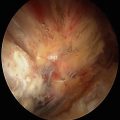
The arthroscope was then placed into the posterior compartment through a posterior trans-tendon portal. Hematoma in the olecranon fossa was evacuated with a shaver in a posterolateral portal. The site of avulsion of the RUHL off the posterior aspect of the lateral epicondyle was visualized just lateral and distal to the olecranon fossa. The arthroscope was easily advanced down the posterolateral gutter due to laxity in the lateral collateral ligament (LCL) complex. The avulsed RUHL was visualized distally in the posterolateral gutter near the level of the radiocapitellar joint (Fig. 26.4). The arthroscope could be advanced from the lateral gutter across the ulnohumeral joint and into the medial gutter, with a positive arthroscopic “drive through sign” of the elbow (Fig. 26.5).
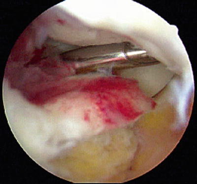
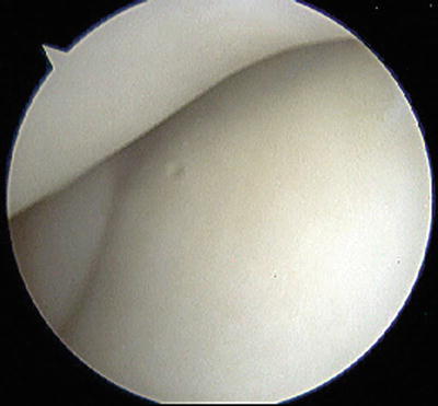

Fig. 26.4
An arthroscopic view of the posterolateral gutter in a left elbow, with the arthroscope in a posterior trans-tendon portal. The shaver is entering the posterior radiocapitellar joint through a lateral soft spot portal. The radial head is visible beyond the shaver. The stump of the RUHL is viewed in the center of the image, avulsed off the posterior aspect of the lateral epicondyle (Published with kind permission. Copyright © Felix H. Savoie, III, MD)

Fig. 26.5
The “drive through sign” of the elbow is demonstrated in this arthroscopic view of the ulnohumeral joint of a left elbow. The arthroscope is in the ulnohumeral joint, viewing from a posterior trans-tendon portal. The articular cartilage of the distal humerus is at the top of the image, with the articular cartilage of the ulna and proximal radioulnar joint at the bottom of the image (Published with kind permission. Copyright © Felix H. Savoie, III, MD)
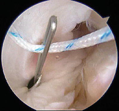
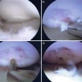
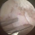
The site of origin of the RUHL was roughened with a shaver, and a double-loaded 2.9 mm suture anchor was inserted percutaneously into the humerus at the origin of the ligament (Fig. 26.6). The sutures were placed down the lateral gutter with a suture retriever. Utilizing a lateral soft spot portal and a percutaneous antegrade suture passer, two mattress sutures were placed in the healthy portion of the ligament. A second suture anchor was placed more distally with a mattress suture placed at the distal aspect of the ligament (Fig. 26.7). The sutures were retrieved and tied, placing the knots deep to the anconeus muscle. As the sutures were tied, tension was restored to the lateral collateral ligament complex, which had the effect of pushing the arthroscope out of the posterolateral gutter. The arthroscopic “drive through sign” could no longer be performed, indicating that adequate tension was restored to the lateral side of the elbow. The arthroscope was then placed back into the anterior compartment. Tension was restored to the annular ligament around the radial head, and the radial head no longer subluxated off the capitellum with forearm supination.
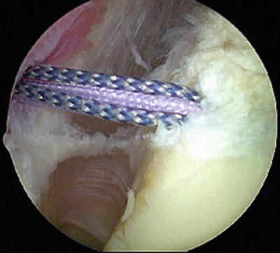
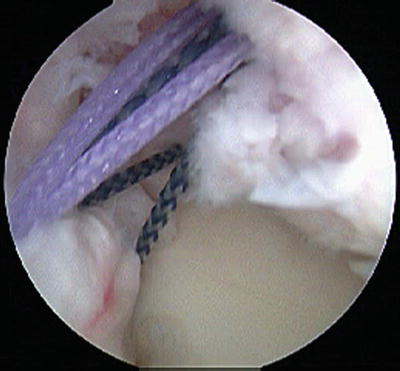

Fig. 26.6
An arthroscopic view of the posterolateral gutter in a left elbow with the arthroscope in a posterior trans-tendon portal. A double-loaded suture anchor has been inserted at the anatomic humeral origin of the RUHL just lateral and distal to the olecranon fossa on the posterior aspect of the lateral epicondyle (Published with kind permission. Copyright © Felix H. Savoie, III, MD)

Fig. 26.7
An arthroscopic view of the posterolateral gutter in a left elbow with the arthroscope in a posterior trans-tendon portal. Mattress sutures have been placed through the healthy portion of the RUHL. The sutures have not yet been tied (Published with kind permission. Copyright © Felix H. Savoie, III, MD)
The patient was splinted for a week and then started physical therapy in a protective brace. The patient returned to play in the brace at 3 weeks, participation in the semifinals and finals. The brace was eliminated at 4 weeks and progress in rehabilitation continued. The patient is currently in his third year of collegiate competition, with no subsequent elbow problems.
Case 2: Chronic PLRI
History/Exam
A 30-year-old, right-hand-dominant, male carpenter presented to the orthopedic clinic complaining of left elbow pain. He had fallen off a roof 3 months prior, landing on his outstretched left arm, and sustained a closed left elbow dislocation. The elbow was reduced in the emergency department, and he was treated conservatively in a hinged elbow brace for 6 weeks. He complained of lateral-sided left elbow pain, feelings of instability, and a palpable clunk with certain activities, especially lifting objects with the left elbow extended.
Physical examination of the left elbow revealed prominence of the radial head and intact skin with no swelling. Range of motion was 10 to 135° of flexion with full rotation. He had tenderness over the radial head and lateral epicondyle, especially posteriorly. He had no gross instability to varus or valgus stress, a positive lateral pivot shift test, and a positive chair push-up test. His wrist exam was normal, and he was neurologically intact distally.
Imaging
Radiographs were obtained with four views of the elbow including AP, lateral, and oblique projections. Radiographs revealed an oval, opaque density on the lateral side of the elbow at the level of the radiocapitellar joint (Fig. 26.8), a concentric reduction of the ulnohumeral joint, and no fractures or subluxations. An MRI arthrogram was ordered with a complete sequence of images; the MRI was ordered with intra-articular contrast, as this was a chronic case. Coronal T2-weighted images are demonstrated in Fig. 26.9a–c, coronal T1-weighted image in Fig. 26.10, sagittal T2-weighted image in Fig. 26.11, and axial T2-weighted image in Fig. 26.12.
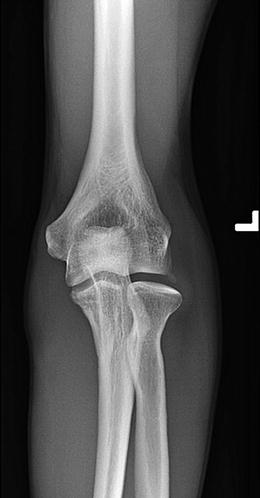
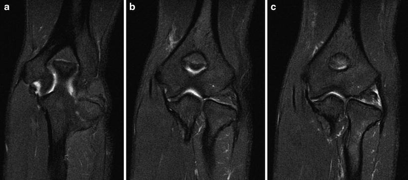
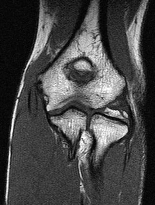
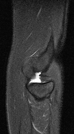
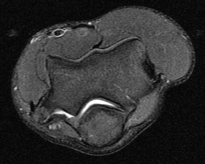

Fig. 26.8
An anteroposterior (AP) radiograph of a left elbow. A small oval density at the level of the radiocapitellar joint represents the avulsed RUHL off the humerus. The ulnohumeral joint is concentrically reduced, and there is slight lateral subluxation of the radial head off the capitellum (Published with kind permission. Copyright © Felix H. Savoie, III, MD)

Fig. 26.9
(a–c) T2-weighted coronal oblique MRI images demonstrate tearing of the RUHL and LCL complex off the posterior aspect of the lateral epicondyle of the humerus. a shows the stump of the RUHL on the right of the image with a void of tissue at the humeral origin of the ligament. b shows heterogeneity in the LCL complex with an intact MUCL on the left side of the image. c shows the stump of the RUHL in the radiocapitellar joint and lateral subluxation of the radial head. The lack of edema in the bone and soft tissues confirms that this is a chronic injury (a–c: Published with kind permission. Copyright © Felix H. Savoie, III, MD)

Fig. 26.10
A T1-weighted coronal oblique MRI image demonstrates heterogeneity of the RUHL and LCL complex on the right side of the image with a small oval of avulsed bone in the stump of the RUHL (Published with kind permission. Copyright © Felix H. Savoie, III, MD)

Fig. 26.11
A T2-weighted sagittal oblique MRI image shows tearing of the RUHL off the posterior aspect of the lateral epicondyle of the humerus, with posterior subluxation of the radial head off the capitellum (Published with kind permission. Copyright © Felix H. Savoie, III, MD)

Fig. 26.12
A T2-weighted axial MRI image shows tearing of the RUHL off the posterior aspect of the lateral epicondyle of the humerus. There is no edema in the soft tissues and no hematoma in the anterior compartment of the elbow joint (Published with kind permission. Copyright © Felix H. Savoie, III, MD)
MRI revealed concentric reduction of the ulnohumeral joint. On the lateral side, there were indistinctness and heterogeneity of the RUHL complex as it neared the humeral attachment (Fig. 26.9a–c) and lateral subluxation of the radial head. Figure 26.9a shows the stump of the RUHL and a void near the humeral attachment. Figure 26.9b shows heterogeneity and thickening of the RUHL and LCL complex, with an intact MUCL on the medial side. Figures 26.9c and 26.10 show the stump of the RUHL in the radiocapitellar joint, with a small avulsed piece of bone on the most proximal aspect of the ligament. The sagittal oblique image (Fig. 26.11) demonstrates the torn RUHL at the level of the radiocapitellar joint. The radial head is subluxated posterior, and the bare area on the posterior aspect of the lateral epicondyle represents the humeral origin of the RUHL. Figure 26.12 also shows avulsion of the RUHL off the posterior aspect of the lateral epicondyle in the axial plane. There is no soft tissue or bony edema on any image sequence, confirming that this represents a remote injury.
The MRI findings of an avulsed RUHL were consistent with the clinical exam of PLRI. Due to the appearance of the RUHL on MRI sequences with a grossly intact ligament avulsed off the humerus with an attached small piece of bone, an arthroscopic repair was offered, with the possible need for open reconstruction. The risks and benefits of the surgery were discussed with the patient. He had failed nonoperative management with clinical instability of the elbow. After an extensive discussion with the patient, he elected to proceed with the proposed surgical intervention.
Arthroscopy
The patient was taken to the operating room and placed in the prone position. Examination under anesthesia demonstrated a positive lateral pivot shift test (Fig. 26.13a, b). A standard diagnostic arthroscopy of the left elbow was completed. As in the first case, laxity of the annular ligament was identified (Fig. 26.14). There was no hematoma in the anterior compartment as this was a case of chronic instability. Rotation of the forearm demonstrated posterolateral subluxation of the radial off the capitellum, indicative of posterolateral rotatory instability.

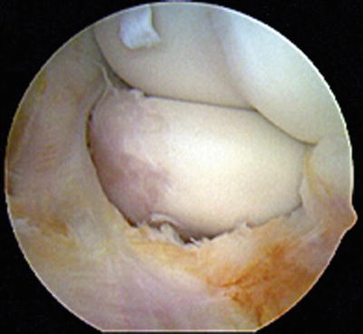

Fig. 26.13
(a, b) Photographs of the lateral pivot shift test in a left elbow. In a, the elbow is reduced. With axial load, valgus force, and supination, the radial head subluxates posterolaterally, and the ulnohumeral joint begins to dislocate (b). The dimple appears proximal to the radial head as the radial head dislocates (a, b: Published with kind permission. Copyright © Felix H. Savoie, III, MD)

Fig. 26.14
An arthroscopic view of the anterior compartment of a left elbow, viewed from a proximal anteromedial viewing portal. Inspection of the radiocapitellar joint shows laxity in the annular ligament and chronic synovitis in the anterior compartment (Published with kind permission. Copyright © Felix H. Savoie, III, MD)
The arthroscope was placed into the posterior compartment through a posterior trans-tendon portal. The arthroscope was again easily advanced down the posterolateral gutter, due to laxity in the LCL complex. The RUHL with attached bone fragment was visualized distally at the level of the radiocapitellar joint. The synovium had a yellowish appearance due to the chronic nature of the injury (Fig. 26.15).
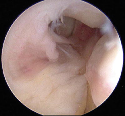

Fig. 26.15
An arthroscopic view of the posterolateral gutter in a left elbow with the arthroscope in a posterior trans-tendon portal. The avulsed RUHL is on the left of the image, sitting in the radiocapitellar joint. The posterior radiocapitellar joint is at the top of the image. The yellowish appearance of the ligament confirms the chronic nature of the injury (Published with kind permission. Copyright © Felix H. Savoie, III, MD)
The site of origin of the RUHL was roughened with a shaver, and a double-loaded 2.9 mm suture anchor was inserted percutaneously into the humerus at the origin of the ligament. Using a percutaneous antegrade suture passer (Fig. 26.16), two mattress sutures were placed into the healthy portion of the ligament, incorporating the lateral capsule (Fig. 26.17). The sutures were retrieved and tied in the same manner as the first case, and tension was restored to the lateral collateral ligament complex.

