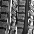
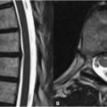
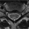
11 Other Vascular Malformations Less common vascular malformations, such as cavernous angiomas (malformations), capillary telangiectasias, and venous angiomas, are more frequently angiographically occult than AVMs. MRI is thus the most sensitive imaging modality for their detection. Cavernous angiomas are the second most common type of vascular malformation after AVMs. They consist of a collection of sinusoidal vascular spaces with no intervening brain parenchyma. Cavernous angiomas are often asymptomatic but may present with hemorrhage or seizures. Supratentorial, subcortical lesions predominate, although lesions occur throughout the CNS and are multiple in up to one-third of cases. Cavernous angiomas are classically described as having a “popcorn-like” heterogeneity on T1WI, T2WI (Figs. 11.1A,B, white arrows), and FLAIR T2WI. Repeated hemorrhage leads to a characteristic thin, well-defined, low SI (on T2WIs), continuous rim of hemosiderin (Figs. 11.1A,B). Areas of hemosiderin deposition are best visualized on T2* weighted GRE T2WI (compare Fig. 11.1B vs Fig. 11.1C
![]()
Stay updated, free articles. Join our Telegram channel

Full access? Get Clinical Tree


