19
 DOUBLE OUTLET RIGHT VENTRICLE
Definition, Spectrum of Disease, and Incidence
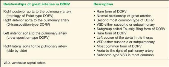
Double outlet right ventricle (DORV) refers to a heterogeneous group of cardiac anomalies where the aorta and pulmonary artery arise primarily from the morphologic right ventricle (Fig. 19-1). The consensus definition of DORV was made deliberately broad by the Congenital Heart Surgery Nomenclature and Database Project, which stated that “DORV is a type of ventriculoarterial connection in which both great vessels arise either entirely or predominantly from the right ventricle” (1). DORV therefore encompasses a family of complex cardiac malformations where both great arteries arise primarily from the morphologic right ventricle but differ with regard to the variable spatial relationship of the great arteries, the location of the ventricular septal defect that is commonly seen with DORV, and the presence or absence of pulmonary and less commonly aortic outflow obstruction. In DORV, four types of anatomic relationships of the aorta to the pulmonary artery at the level of the semilunar valves have been described (2) and include a right posterior aorta, a right anterior aorta, a left anterior aorta, and a right lateral aorta (Table 19-1). The ventricular septal defect (VSD) that is commonly associated with DORV has been described in four anatomic locations: subaortic type; subpulmonary type; subaortic and subpulmonary type (also called doubly committed); and remote type, nonrelated to both arteries (Table 19-2). The exact subtype of DORV may be hard to characterize prenatally as the position of the VSD is difficult to establish with accuracy in fetal echocardiography.
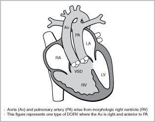
Figure 19-1. Double outlet right ventricle (DORV). LV, left ventricle; LA, left atrium; RA, right atrium; VSD, ventricular septal defect.
| TABLE 19-1 | Anatomic Relationships of the Great Arteries at the Semilunar Valves in Double Outlet Right Ventricle (DORV) |
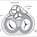
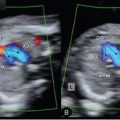
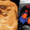
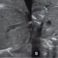
DORV occurs in about 1% to 1.5% of children born with congenital heart disease and has an incidence of approximately 0.09 per 1000 live births (3). It is found equally in boys and girls. DORV is more common in fetal series and has been reported in up to 6% of congenital heart disease (4). In fetal series, DORV is reported either as a separate entity or within the group of conotruncal anomalies (4–11). The prevalence of DORV is increased in diabetic pregnancies (12). Figures 19-2 and 19-3 represent anatomic specimens of two types of DORV in fetal hearts.
Ultrasound Findings
Related posts:
Stay updated, free articles. Join our Telegram channel

Full access? Get Clinical Tree


