Presentation and Presenting Images
( ▶ Fig. 70.1, ▶ Fig. 70.2, ▶ Fig. 70.3, ▶ Fig. 70.4, ▶ Fig. 70.5, ▶ Fig. 70.6, ▶ Fig. 70.7)
A 38-year-old female with an abnormal baseline mammogram at an outside institution presents for diagnostic mammography.
70.2 Key Images
( ▶ Fig. 70.8, ▶ Fig. 70.9, ▶ Fig. 70.10, ▶ Fig. 70.11, ▶ Fig. 70.12, ▶ Fig. 70.13, ▶ Fig. 70.14)
70.2.1 Breast Tissue Density
There are scattered areas of fibroglandular density.
70.2.2 Imaging Findings
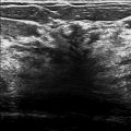
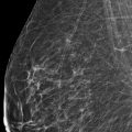
The imaging of the right breast is normal (not shown). The left breast demonstrates a 1-cm isodense oval mass in the posterior depth of the left breast at the 6 to 7 o’clock location, 10 cm from the nipple ( ▶ Fig. 70.8, ▶ Fig. 70.9, ▶ Fig. 70.10). A fatty hilum is suggested on the lateromedial (LM) spot-compression digital breast tomosynthesis (DBT) slice 37 of 41 ( ▶ Fig. 70.12 and ▶ Fig. 70.14) and the craniocaudal (CC) spot-compression DBT slice 19 of 69 ( ▶ Fig. 70.11 and ▶ Fig. 70.13). A skin marker (arrow) denotes an unrelated skin lesion.
70.3 BI-RADS Classification and Action
Category 2: Benign; resume routine mammography
70.4 Differential Diagnosis
Intramammary lymph node: Although this is an uncommon location for an intramammary lymph node, the fatty hilum helps to make the diagnosis.
Cyst: Cysts are often multiple, usually bilateral, and may be painful and fluctuate in size. They are most common in 30- to 50-year-old patients. The fatty hilum makes a cyst an unlikely possibility.
Fibroadenoma: Fibroadenomas are the most common benign masses in young women. They are multiple in 10 to 15% of patients. The fatty hilum makes a fibroadenoma an unlikely possibility.
70.5 Essential Facts
A normal intramammary lymph node is a well-circumscribed, slightly lobulated mass which is most instances contains a radiolucent cleft, which represents fat in the hilum of the node.
A fatty hilum is suggested with the mass in this case; however, this mass is not in the typical location for an intramammary lymph node—the upper outer quadrant. Meyer et al (1993) suggests that no further work-up is necessary for a typical-appearing lymph node in an abnormal location.
DBT evaluation demonstrating the fatty hilum helps to make this diagnosis.
70.6 Management and Digital Breast Tomosynthesis Principles
DBT of the entire breast or spot compression (as in this example) may be performed. All views performed with conventional two-dimensional (2D) mammography may be performed with DBT except spot-magnification views.
Distant metastases are the major cause of mortality and morbidity in breast cancer patients. Axillary node metastases have proven to be the most important prognostic indicator, and are significantly associated with decreased disease-free survival and overall survival time after diagnosis. Intramammary nodes are seen on all breast imaging modalities, but the clinical significance of a metastatic intramammary node is not well established.
The preoperative detection and accurate characterization of intramammary lymph nodes is vital to stage and plan treatment for these patients, particularly for patients with axillary-lymph-node–negative disease. A metastatic intramammary lymph node upgrades the disease and warrants further axillary dissection at the time of surgery and these patients may be candidates for neoadjuvant systemic therapy.
70.7 Further Reading
[1] Hogan BV, Peter MB, Shenoy H, Horgan K, Shaaban A. Intramammary lymph node metastasis predicts poorer survival in breast cancer patients. Surg Oncol. 2010; 19(1): 11‐16 PubMed
[2] Mahajan A, Udare A, Shet T, Juvekar S, Thakur M. Diagnosis of a malignant intramammary node retrospectively aided by mastectomy specimen MRI-Is the search worth it? A case report and review of current literature. Korean J Radiol. 2013; 14(4): 576‐580 PubMed
[3] Meyer JE, Ferraro FA, Frenna TH, DiPiro PJ, Denison CM. Mammographic appearance of normal intramammary lymph nodes in an atypical location. AJR Am J Roentgenol. 1993; 161(4): 779‐780 PubMed

Fig. 70.1 Left craniocaudal (LCC) mammogram.
Stay updated, free articles. Join our Telegram channel

Full access? Get Clinical Tree


