67
Overview of Liver Lesions
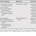
Hemangioma
Demographics
Classic Imaging Appearance
Differentiating Features
Hemangioma versus Hypervascular Metastasis or Hepatocellular Carcinoma
Cholangiocarcinoma versus Hemangioma
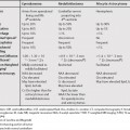
Focal Nodular Hyperplasia
Demographics
Classic Imaging Features
Differentiating Features
Focal Nodular Hyperplasia versus Adenoma
Focal Nodular Hyperplasia versus Fibrolamellar Hepatocellular Carcinoma
Hepatic Adenoma
Demographics
Classic Imaging Features
Differentiating Features
Adenoma versus Focal Nodular Hyperplasia
Hepatocellular Carcinoma
Demographics
Classic Imaging Appearance
Differentiating Features
HCC versus Regenerating or Dysplastic Nodule
Related posts:

Stay updated, free articles. Join our Telegram channel

Full access? Get Clinical Tree


