Fig. 3.1
A bird’s-eye view of the HIMAC facility A unique double-synchrotron ring heavy-ion accelerator system dedicated for medical use was designed and constructed. It consists of two ion sources, an RFQ (radio frequency quadrupole) linear accelerator (linac), an Alvarez linear accelerator (linac), two synchrotron rings, a high-energy beam transport system, and an irradiation system
Table 3.1
Specifications of the HIMAC
Ion: H–Ar | |
Max energy | 100–800 MeV/u |
Treatment room [3] | |
Fixed vertical | Room A |
Fixed horizontal | Room C |
Fixed vertical and horizontal | Room B |
Accelerated energy | |
Vertical beam Horizontal beam | 140 or 290 or 350 or 400 MeV/u 140 or 290 or 400 or 430 MeV/u |
Range of carbon ion beam in water | |
140 MeV/u | 5 cm |
290 MeV/u | 15 cm |
350 MeV/u | 20 cm |
400 MeV/u | 25 cm |
430 MeV/u | 30 cm |
Maximum field size | 15 cm by 15 cm |
In order to conform the dose to a specific target, a passive beam delivery technique has been employed. The beam lines in the treatment room are equipped with a pair of wobbler magnets, beam scatterers, ridge filters, multi-leaf collimators, and the ability to administer a compensation bolus. An appropriately designed ridge filter, which corresponds to and determines the size of the spread out of Bragg peak (SOBP), is chosen to avoid unnecessary dosing of normal tissues along the beam path in each port. Twelve different size ridge filters were made to cover thicknesses of 2–15 cm.
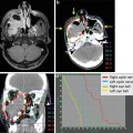
The patients are positioned in customized cradles and immobilized with a low-temperature thermoplastic cast. A set of CT images are taken for treatment planning with the immobilization devices in place. Respiratory gating for both the CT acquisition and therapy is performed when indicated [3]. A three-dimensional treatment planning is performed using our original HIPLAN software program, which was developed for carbon ion radiotherapy CIRT [4]. A margin of 5 mm is usually added to the clinical target volume to create the planning target volume. The dose is calculated for the target volume and any nearby critical structures and expressed in Gray-equivalents (GyE = carbon physical dose in Gray × Relative Biological Effectiveness {RBE}).
Radiobiological studies were carried out in mice and in five human cell lines cultured in vitro to estimate RBE values relative to megavoltage photons. Irrespective of the size of the SOBP, the RBE value of carbon ions was estimated to be 3.0 at the distal part of the SOBP, and ridge filters were designed to produce a physical dose gradient of the SOBP so that the biological effect along the SOBP became uniform. This was based on the biologic response of human salivary gland tumor cells at a 10 % survival level. The biological response flatness along the SOBP was checked by measuring the physical dose distributions and dose-averaged LET, which were in satisfactory agreement with the calculated results [5].
Carbon ion radiotherapy (C-ion RT) is given once daily, 4 day/week (Tuesday to Friday). During every treatment session, the patient’s position is verified with a computer-aided online positioning system.
A new beam delivery with active scanning was started in May 2011, and we have since treated more than 200 patients with fixed targets.
3.3 Clinical Trials at the NIRS
From June 1994 until March 2013, a total of 70 protocols were conducted in an attempt to determine the optimal dose-fractionation and irradiation method for the treatment of specific diseases. The HIMAC passive beam delivery system has been showing reliable and stable performance for the last 19 years, and a total of more than 7,000 patients had been registered for treatment, and the C-ion RT was approved as a clinical practice in 2003. The number of patients treated as part of clinical practice as of June 2013 has been more than 4,000 at the NIRS (Fig. 3.2). The categories of disease that can be treated in routine clinical practice include lung cancer, prostate cancer, head and neck cancer, skull base tumors, ocular melanoma, bone and soft tissue sarcoma, liver cancer, pelvic recurrences of rectal cancer, pancreatic cancer, uterine cervical cancer, and re-irradiation after conventional radiotherapy, among others. The number of patients has increased every year, and the facility has reached a capacity permitting more than 800 patients to be treated each year (Fig. 3.3).
Fig. 3.2
The number of patients treated using carbon ion radiotherapy from June 1994 to March 2013 at the NIRS. A total of 7,349 patients had received carbon therapy at the NIRS as of March 2013. Prostate cancer, sarcoma, head and neck cancer, lung cancer, liver cancer, postoperative local recurrence of rectal cancer, and pancreatic cancer were the most frequently treated tumors at the NIRS. More than 4,000 patients have received carbon ion radiotherapy as part of clinical practice
Fig. 3.3


Related posts:
Stay updated, free articles. Join our Telegram channel
Full access? Get Clinical Tree
