Presentation and Presenting Images
( ▶ Fig. 80.1, ▶ Fig. 80.2, ▶ Fig. 80.3, ▶ Fig. 80.4, ▶ Fig. 80.5, ▶ Fig. 80.6)
A 31-year-old female with bilateral palpable breast masses presents for mammographic evaluation.
80.2 Key Images
( ▶ Fig. 80.7, ▶ Fig. 80.8, ▶ Fig. 80.9, ▶ Fig. 80.10, ▶ Fig. 80.11, ▶ Fig. 80.12, ▶ Fig. 80.13, ▶ Fig. 80.14)
80.2.1 Breast Tissue Density
The breasts are heterogeneously dense, which may obscure small masses.
80.2.2 Imaging Findings
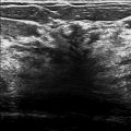
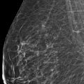
The patient has palpable abnormalities denoted by triangular markers (arrows in ▶ Fig. 80.7, ▶ Fig. 80.8, ▶ Fig. 80.9, and ▶ Fig. 80.10) in the upper outer quadrants of both breasts (1 o’clock location in left breast and 11 o’clock location in right breast). The triangular markers are not well seen on the right mediolateral oblique (MLO) and right lateromedial (LM) views ( ▶ Fig. 80.3 and ▶ Fig. 80.5). Historically the patient has solid masses in the right breast at the 3 o’clock location and the 10 o’clock location. These have been previously biopsied; however, biopsy clips were not placed at the time of the outside biopsies. The right 10 o’clock mass (circle in ▶ Fig. 80.13 and ▶ Fig. 80.14) is a oval hypoechoic mass, which is a fibroadenomatous change, and the right 3 o’clock mass (circle in ▶ Fig. 80.11 and ▶ Fig. 80.12) is an oval mixed echoic mass, which is a fibroadenoma. There are no correlates on conventional mammography or digital breast tomosynthesis for the current bilateral palpable abnormalities or the known right breast masses. The ultrasound of the current bilateral palpable areas (triangle markers) were consistent with benign breast parenchyma (not shown).
80.3 BI-RADS Classification and Action
Category 2: Benign
80.4 Differential Diagnosis
Normal breast tissue: Normal breast tissue is a common cause of palpable breast masses.
Cysts: Cysts are often multiple, usually bilateral, and may be palpable and/or painful and fluctuate in size. They are most common in 30- to 50-year-old patients.
Fibroadenomas: Fibroadenomas are the most common benign masses in young women. They are multiple in 10 to 15% of patients with them. Fibroadenomas may be palpable.
80.5 Essential Facts
Palpable breast masses are common and usually benign, but accurate evaluation and prompt diagnosis are necessary to rule out malignancy.
Additional supporting imaging or clinical information is helpful in diagnosis and management.
In this case, there are no imaging correlates noted on conventional mammography or DBT for the bilateral palpable abnormalities or the known right breast masses.
The biopsy-proven benign masses at the 3 o’clock and 10 o’clock positions of the right breast were unchanged in size by ultrasound.
80.6 Management and Digital Breast Tomosynthesis Principles
There will be tomosynthesis-occult lesions. Further research is needed to determine which lesions are more likely to be tomosynthesis occult.
There is a paucity of literature regarding lesions that are occult on tomosynthesis.
Palpable findings without a mammographic or tomosynthesis correlate must be evaluated with ultrasound.
The work-up of a palpable finding cannot stop with the absence of a mammographic or tomosynthesis finding. The palpable mass may be mammographically or tomosynthesis occult, as in this case. Palpable masses must be evaluated completely with ultrasound. If there is no ultrasound correlate, further work-up should be based upon the clinical suspicion.
80.7 Further Reading
[1] Klein S. Evaluation of palpable breast masses. Am Fam Physician. 2005; 71(9): 1731‐1738 PubMed
[2] Kopans DB. Digital breast tomosynthesis: a better mammogram. Radiology. 2013; 267(3): 968‐969 PubMed

Fig. 80.1 Right craniocaudal (RCC) mammogram.
Stay updated, free articles. Join our Telegram channel

Full access? Get Clinical Tree


