18 Pancreatic Arteries
K.I. Ringe, S. Meyer
The arterial supply of the pancreas is highly variable, but follows a general pattern.1–22
The head of the pancreas is supplied by anastomosing branches from the celiac trunk and the superior mesenteric artery. These arteries form arcades in front of and behind the head of the pancreas on the vertical axis.
The body of the pancreas is supplied by arteries running in the anterior and posterior arcades of the head of the pancreas; these arteries vary not only in number but also in origin. The anterior superior pancreaticoduodenal artery consistently represents the terminal branch of the gastroduodenal artery. Both inferior origins of the anterior and posterior arcades leave the superior mesenteric artery as a short common trunk (Fig. 18.5); alternative origins for the anterior arcade are shown in Fig. 18.6, Fig. 18.7, and Fig. 18.8.23 The superior origin of the posterior arcade varies more often (Fig. 18.11, Fig. 18.12, Fig. 18.13, and Fig. 18.14). The transverse pancreatic artery to the body of the pancreas is connected with the posterior pancreatic and the large pancreatic arteries. The transverse pancreatic artery is often referred to as the inferior pancreatic artery or, less often, the superior pancreatic artery. This might partly explain the considerable discrepancy in the literature on the frequency of this artery. In addition to the origins shown in Fig. 18.20, Fig. 18.21, and Fig. 18.22, other origins, such as the common hepatic artery, the gastroduodenal artery, and the posterior arcade, have been described.
The cauda of the pancreas is supplied by numerous branches from the splenic artery,5,20 which are not shown in the figures.
In selective pancreatic angiography, the number of arteries and branches identified largely depends on the position of the catheter and the direction of the blood flow in the different anastomosing arteries around or within the pancreas. Fewer pancreatic arteries are detected than in anatomical studies. The large pancreatic artery (arteria pancreatica magna) has not been depicted in detail in the figures in this chapter.
18.1 Pancreatic Arteries as Shown in Textbooks
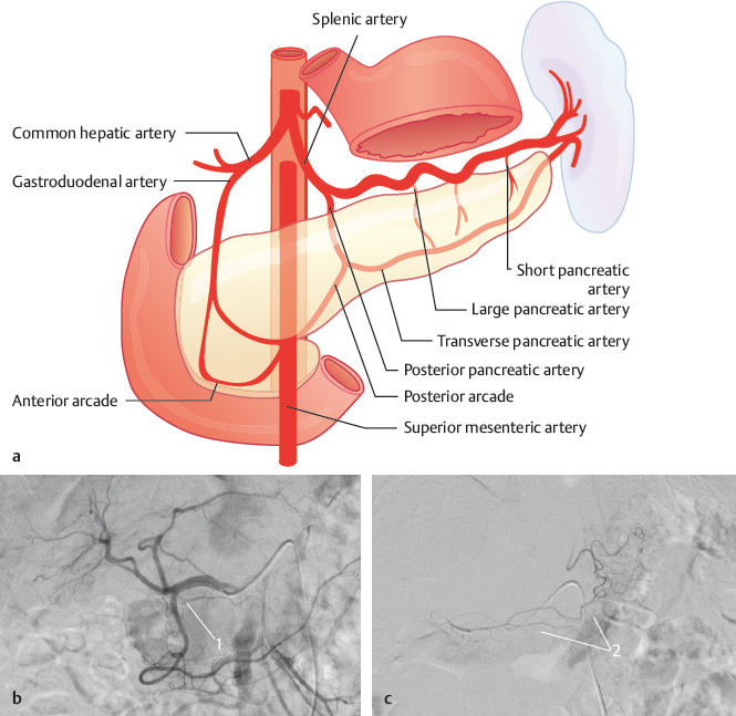
Fig. 18.1 “Normal” blood supply of the pancreas. Schematic (a) and angiography (b,c) with a catheter in the common hepatic artery (b) and a microcatheter in the anterior arcade (c). 1 Anterior arcade; 2 pancreas.
18.2 Number of Arterial Arcades in Front of the Head of the Pancreas
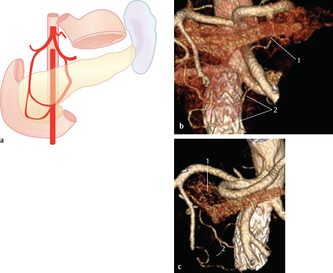
Fig. 18.2 Two arcades (79%). Schematic (a) and oblique VR CT, coronal (b) and sagittal (c) view. 1 Pancreas; 2 two arcades.

Fig. 18.3 Three arcades (16%). Schematic.
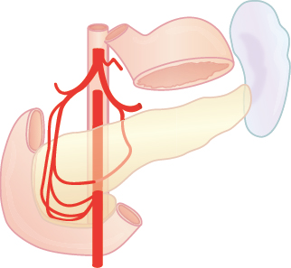
Fig. 18.4 Four arcades (5%). Schematic.
18.3 Inferior Origin of the Anterior Arcades

Fig. 18.5 Inferior origins of both the anterior and the posterior arcades leave the superior mesenteric artery as a short common trunk (65%). Schematic.

Fig. 18.6 Inferior origin from the right side of the stem of the superior mesenteric artery (8%). Schematic (a) and coronal VR CT (b). 1 Anterior arcade inferior origin; 2 superior mesenteric artery.

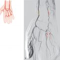
Fig. 18.7 Inferior origin from the first jejunal artery (15%). Schematic (a) and angiography (b) with a catheter in the superior mesenteric artery. 1 Anterior arcade; 2 first jejunal artery.
Related posts:


Stay updated, free articles. Join our Telegram channel

Full access? Get Clinical Tree


