, Leonid Tiutin2 and Thomas Schwarz3
(1)
Russian Research Center for Radiology and Surgery, St. Petersburg, Russia
(2)
Department of Radiology and Nuclear Medicine, Russian Research Center for Radiology, St. Petersburg, Russia
(3)
Department of Nuclear Medicine Division of Radiology, Medical University Graz, Graz, Austria
Abstract
Epidemiological studies of recent years have registered a steadfast growth in the number of pancreatic cancer cases. In the Russian Federation this number grows by 1.2% annually, and the increment of pancreatic cancer cases for recent years has been 11%. The share of pancreatic cancer in oncological mortality makes up 5% (Aksel et al. 2001).
Epidemiological studies of recent years have registered a steadfast growth in the number of pancreatic cancer cases. In the Russian Federation this number grows by 1.2% annually, and the increment of pancreatic cancer cases for recent years has been 11%. The share of pancreatic cancer in oncological mortality makes up 5% (Aksel et al. 2001).
Smoking, excessive consumption of meat products and fat, use of alcohol in big doses and hard manual labor are the essential risk factors for pancreatic cancer. There are cases of occurrence of a malignant neoplasm with antecedent pancreatic diabetes. Consequently persons older than 50 years of age who have a sudden development of diabetes, especially without hereditary load, should be considered to belong to the group of risk. Primary chronic pancreatitis may be also a background disease for development of pancreatic cancer due to dysplastic, metaplastic and disregeneratory changes of epithelium of the pancreas (Kazyulin and Kucheryavyi 2005; Kubyshkin and Vishnevsky 2003).
The clinical picture of pancreatic cancer most often develops gradually. On average, 3–4 months pass between the start of clinical manifestations and diagnosis. Among the initial manifestations of pancreatic cancer there are discomfort in the upper part of the abdomen (sensation of heaviness and repletion, especially after meal), pain, loss of weight, lowering of the appetite and nausea. The most apparent symptom appearing in the late stages of the disease is obturative jaundice, whose frequency and intensity are influenced by tumor localization and size, as well as by its relation to biliary ducts. In 70% of cases, jaundice is preceded by different symptoms caused by background diseases and complications of tumor growth. Therefore, several clinical forms of pancreatic cancer during the pre-jaundice period are distinguished: pancreatitis-like, diabetoid, cholangitic and gastritis-like. The duration of the period without jaundice is from 1 to 6 months. Tumor invasion to the duodenum is characterized by symptoms of gastroduodenal obstruction, intestinal hemorrhage and cholangitis (Maev 2004 and Putov 2008).
10.1 Pathologic Anatomy and Mechanisms of Metastases
Growth of malignant tumor of the pancreas occurs by way of cancer infiltration of the inter-tissue space and lymphatic sheets lying inside glandular lobules, as well as through connective-tissue spaces surrounding ducts, vessels and nerves of the pancreas. Its further expansion leads to the invasion of the capsule of the pancreas and to infiltration of retroperitoneal cellular tissue in which the portal vein and its affluxes are situated. Depending on the location of malignant neoplasm in the pancreas, it is accepted to distinguish cancer of the head, body and tail of the pancreas as well as diffuse or total lesion of the organ. The frequency of cancer of the head of the pancreas is 58–75%, that of the body of pancreas is 12–23% and that of the tail of the pancreas is 5–7%; the frequency of diffuse lesion being 1–6%. The dimensions of the tumor node range from several millimeters to 12–14 cm. Tumors of the head and tail of the pancreas are usually more massive than those of the corpus.
In 80–90% of cases it is adenocarcinoma in various degrees of differentiation that is the morphologic norm of ductal cancer of the pancreas. Rarer are planocellular, glandular-planocellular, anaplastic, giant-cell cancers and carcinosarcoma. Acinic-cell (parenchymatous) cancer microscopically belongs to alveolar tumors and occurs most often in the area of the body and tail of the pancreas.
Tumor dissemination in pancreatic cancer occurs in three ways: lymphogenous, hematogenous and by implantation. Tumor metastasis through lymph collectors takes place in several stages:
Stage 1 – lesion of pancreatoduodenal lymph nodes
Stage 2 – lesion of retropyloric and hepatoduodenal lymph nodes
Stage 3 – lesion of celiac and superior mesenteric lymph nodes
Stage 4 – lesion of retroperitoneal (most frequently paraaortal) lymph nodes
In the cases of hematogenous and implantation ways of tumor dissemination, distant metastases are observed in the liver, lungs, kidneys, bone system and peritoneum.
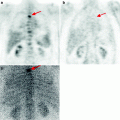
10.2 Methods of Diagnosis
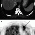
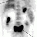
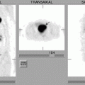
Presently US is the first stage of the diagnostic algorithm in examining patients with solid pancreatic masses. The diagnostic accuracy of the method in detecting pancreatic cancer ranges within large limits and varies from 48% to 86%. The sensitivity of US in the diagnosis of invasion of great blood vessels corresponded to 43% and in determining tumor resectability was 30.5% (Arablinsky et al. 1992; Kotlyarov 1999; Casadet et al. 2002). The appearance of multi-layer spiral CT has considerably increased the possibilities for radiodiagnosis. When combined with intravenous or intraarterial (CT arterioportography) contrast, the accuracy of the method may reach 82–93% (Bronstein et al. 2004).
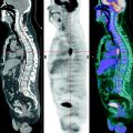
MRI is one of the most efficient radiological methods of examining the duodenal area. Use of dynamic contrast-enhanced MRI permits the performance in many cases of the differential diagnosis of pseudotumorous pancreatitis and tumor lesion of the pancreas. The introduction of MR aortoportography and venoportography has considerably increased the information value of MR examinations, as well as MR cholangiopancreatography, which permits the visualization of biliary tracts and the duct of Wirsung. Complex application of all the mentioned MR techniques permits an increase in the diagnostic accuracy of the method in pancreatic cancer of up to 88–97% (Amthauer et al. 2002).
Much importance in the diagnosis of pancreatic cancer is still attached to endoscopic X-ray cholangiopancreatography. When implementing it, one can carry out intake of pancreatic juice, and obtain material by brush-biopsy for cytological study with rather high efficiency. However, changes in pancreaticograms in patients with chronic pancreatitis are detected in 72–76% and in pancreatic cancer patients only in 24% of cases; if tumor is localized in the uncus they are detected in no more than 10–15% of patients. In 10% of cases, pancreaticograms in the event of pancreatic cancer resemble those for chronic pancreatitis (Revyakin et al. 1996; Schofl and Haefner 2003).
It should be noted that despite the wide spectrum of radiological methods of examination, the problem of differential diagnosis of pancreatic cancer and pseudotumorous pancreatitis cannot be considered as resolved. In many cases the similarity of radiological symptoms in both diseases does not permit to differentiate between tumor lesion of the pancreas and benign focal hyperplasia, which finally results in rather low specificity of the obtained data. At the same time, the adequacy of treatment, prognosis and life quality of patients with solid masses of the pancreas directly depend on timely and accurate diagnosis.
Another important problem is the diagnosis of metastatic lesion of lymph nodes in pancreatic cancer. In US and CT examinations, the enlargement of lymph nodes is the most frequent (and in many cases the only) sign of their tumor affection. Meanwhile, changes in size of a lymph node do not always mark it as a malignant lesion. Cases are known when the diameter of a lymph node in pancreatic cancer corresponded to 40 mm according to US and CT. However, in a morphological study of biopsy material, signs of reactive hyperplasia were detected. The main cause of enlargement of a lymph node in pancreatic cancer lies in marked inflammatory changes in glandular tissue, which usually accompany a malignant process.
Related posts:
Stay updated, free articles. Join our Telegram channel

Full access? Get Clinical Tree


