Chapter Outline
Types of Pancreas Transplantation
Pancreas Transplant Imaging: Normal Appearances
Pancreas Transplant Imaging: Postoperative Complications
Pancreas transplantation offers patients with severe type 1 diabetes mellitus the ability to restore normoglycemia and to halt or reverse the progression of diabetic complications, such as nephropathy, retinopathy, and vasculopathy. The standard procedure is transplantation of a whole-organ cadaveric pancreas while the recipient’s native pancreas is left in place. Surgical techniques are varied and have evolved during the last four decades. Despite improvements during this period in both patient and graft survival rates, graft loss occurs with high prevalence. Early detection of graft-related complications is fundamental for graft survival and often relies on multiple imaging modalities. Imaging after pancreas transplantation poses a special challenge to the radiologist because it requires knowledge of the transplantation procedure, the complex postsurgical anatomy, and a wide spectrum of postoperative complications. Ultrasound (US), computed tomography (CT), and magnetic resonance imaging (MRI) offer specific advantages and limitations in this setting, and a multimodality approach is often required to optimally evaluate the pancreas transplant. In this chapter, we review the spectrum of surgical procedures, postoperative anatomy, and complications of pancreas transplantation with emphasis on the normal and abnormal findings seen at US, CT, and MRI.
Types of Pancreas Transplantation
According to the International Pancreas Transplant Registry, more than 26,000 pancreas transplantations have been performed in the United States since 1988; 1042 were performed in 2012. Three types of pancreas transplantation procedures are typically performed. Most commonly, the pancreas is transplanted from a deceased donor together with a kidney transplantation occurring at the same time (simultaneous pancreas-kidney [SPK] transplantation); this accounts for 75% of pancreas transplants performed in the United States today. Less commonly (18% of cases), a patient receives a pancreas transplant after having already undergone successful kidney transplantation from a live or a deceased donor (pancreas after kidney [PAK]) transplantation). Pancreas transplantation alone (PTA) is performed least commonly, accounting for 7% of cases.
SPK transplants allow patients to be free of both insulin therapy and dialysis and result in higher 1-year graft survival rates (86%) than PAK or PTA transplants (80% and 78%, respectively). This difference persists in the long term, with SPK transplants demonstrating a graft function rate of 45% at 20 years compared with 16% for PAK and 12% for PTA transplants. The advantages of pancreas transplantation must be balanced against short-term surgical complications and long-term immunosuppressive regimens. However, with recent advances in surgical technique, human leukocyte antigen matching, and immunosuppressive regimens, all types of pancreas transplants have shown significant improvements in both patient and graft survival rates during the last two decades. Living donor pancreas transplants account for a small minority of cases (0.5%) and are not discussed.
Pancreas Transplant Anatomy
Pancreas Procurement
The pancreas allograft is harvested with its vascular support and a segment of the donor duodenum containing the ampulla of Vater. The donor’s iliac artery bifurcation is also procured for reconstruction of the arterial conduit. Surgical techniques have evolved over time to deal with arterial inflow, venous outflow (endocrine secretions), and pancreatic duct exocrine drainage, with the most variability in the venous and duodenal attachments.
Arterial Supply
The pancreas transplant receives arterial inflow from two sources: the donor superior mesenteric artery, which supplies the head through the inferior pancreaticoduodenal artery; and the donor splenic artery, which supplies the body and tail. The internal and external iliac arteries of the donor iliac bifurcation are attached in an end-to-end fashion to the donor superior mesenteric artery and splenic arteries, forming a Y graft ( Figs. 100-1 and 100-2 ). The common iliac portion is then anastomosed to the recipient common iliac artery or external iliac artery, with a variable graft length depending on the venous and duodenal attachments.


Venous Drainage
Venous outflow from the graft may be drained to the recipient’s portal or systemic venous system. Pancreas transplantations were originally performed with systemic venous drainage. However, pancreatic endocrine secretions contained in the venous outflow raised concerns about systemic hyperinsulinemia causing accelerated atherosclerosis, hypertension, and hypercholesterolemia and resulted in a change to the more physiologic state of portal venous drainage. This is accomplished by grafting the donor portal vein, which functions as the main graft vein, to the recipient superior mesenteric vein (see Figs. 100-1 and 100-2A ). Systemic venous drainage involves an anastomosis of the graft vein to the recipient iliac vein (see Fig. 100-2B ) or, rarely, to the inferior vena cava. These techniques are currently equivalent in terms of patient and short-term graft survival, and although systemic drainage is currently predominant, portal venous drainage is still widely used.
Exocrine Secretions
Pancreatic exocrine secretions drain into the donor duodenal stump that is harvested with the pancreas. This can be attached side-to-side to the recipient’s small bowel for enteric drainage or to the recipient’s bladder. Enteric exocrine drainage is used most commonly (91% of SPK, 89% of PAK, 85% of PTA), with or without the creation of a Roux-en-Y loop (see Fig. 100-2A ). Enterically drained transplants are usually located in the midabdomen to the right of midline, with the head of the pancreas situated cranially for portal venous drainage or caudally for systemic venous drainage. Bladder drainage of exocrine secretions is achieved by an anastomosis between the donor duodenal stump and the superior aspect of the bladder (see Fig. 100-2B ). Bladder-drained allografts are usually located in the right pelvis, superior to the bladder, with the head of the pancreas directed caudally. One advantage of bladder drainage is that urinary amylase may be used to monitor graft function. However, in many centers, enteric drainage is preferred to bladder drainage because it is associated with fewer metabolic complications and lower rates of hematuria and recurrent urinary infections. Enteric conversion may be successfully performed after initial bladder drainage; up to 25% of patients undergo this procedure.
Pancreas Transplant Imaging: Normal Appearances
Ultrasound
US is the most commonly used imaging modality to evaluate the transplanted pancreas; it is routinely used in its initial evaluation. Gray-scale imaging can demonstrate the allograft and peripancreatic fluid collections; color and duplex Doppler imaging can document perfusion and evaluate the arterial and venous vasculature. The well-known advantages of US include availability, repeatability, and portability for ill and unstable patients in the postoperative setting. US can also be used to guide percutaneous biopsy of the transplant pancreas. Sonographic evaluation is, however, operator dependent, and anatomic challenges, such as obscuration by bowel, are not uncommon. Unless it is abnormally dilated, the duodenal component most often cannot be separately evaluated. When direct visualization of the transplant pancreas is difficult, color and power Doppler imaging can help in identifying parenchymal and graft vessel flow, thereby localizing the pancreas.
The normal pancreas transplant is a homogeneous soft tissue structure hypoechoic relative to surrounding mesenteric fat on gray-scale US ( Fig. 100-3A ). Because it lacks a capsule, its borders may be indistinct, and it may be difficult to distinguish the allograft from adjacent structures. Color and power Doppler are essential to demonstrate pancreas transplant perfusion and vascular anatomy; the Y arterial graft, graft vein, and splenic artery and vein are usually visible ( Fig. 100-3B -D). Arterial waveforms normally show a rapid systolic upstroke and continuous diastolic blood flow, whereas venous structures demonstrate a monophasic waveform within an anechoic lumen ( Fig. 100-3E ). Arterial resistive indices are variable within the graft and are not useful in distinguishing normal from abnormal grafts.

Computed Tomography
CT is most often performed when there is concern for abdominal infection, bowel complication, or pancreatitis or its complications. It is better suited to evaluate fluid collections and bowel complications because of its wider field of view and intrinsic properties. CT can reliably demonstrate the pancreas, the duodenal stump, and the recipient small bowel, which can be useful when the transplant cannot be visualized sonographically because of overlying bowel gas. This may be particularly helpful in guiding percutaneous biopsy.
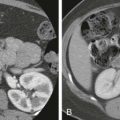
Positive enteric contrast agents should be used to differentiate bowel from fluid collections and from the transplant. Because most allografts are SPK transplants, CT is usually performed without intravenous contrast material to avoid further renal injury in patients with impaired renal function. This limits evaluation of the vasculature and parenchymal enhancement. On unenhanced CT, the allograft appears as a homogeneous soft tissue structure that may be isodense to and difficult to distinguish from unopacified and nondistended bowel, although surgical staples on either side of the duodenal stump are helpful for localization ( Fig. 100-4A ). The donor duodenum is often collapsed and thick walled but can be misinterpreted as a fluid collection when it is distended. It inconsistently opacifies with oral contrast material even when the adjacent jejunum is contrast filled. Contrast-enhanced CT, especially with multiplanar reformations, can demonstrate the vascular anatomy and parenchymal enhancement well ( Fig. 100-4B, C ).

Magnetic Resonance Imaging and Angiography
MRI and magnetic resonance angiography (MRA) are used less frequently in the evaluation of the transplanted pancreas. They are most commonly performed to confirm the diagnosis of a vascular complication, after an abnormal or nondiagnostic US or CT study. High-resolution, three-dimensional (3D), contrast-enhanced MRA provides an accurate diagnostic technique for evaluation of the arterial and venous anatomy of the pancreas transplant. Unenhanced MRI sequences readily separate the pancreatic allograft from adjacent structures and are superior to CT without intravenous contrast material. MR is particularly useful when US is limited by overlying bowel gas or by body habitus. Given the increased risk for nephrogenic systemic fibrosis in patients with end-stage renal disease, the risk/benefit ratio of intravenous gadolinium-based contrast agents should be carefully assessed. Unenhanced MRA is unlikely to perform as well as contrast-enhanced MRA, but it has not been formally evaluated in this setting.
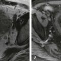
On T1-weighted images, the pancreatic parenchyma is homogeneous and hyperintense relative to the liver. On T2-weighted images, the normal pancreas allograft will show signal intensity between fluid and muscle ( Fig. 100-5A ). T2-weighted images are most sensitive to abnormalities of the pancreatic transplant because the majority of pathologic processes increase glandular water content. We typically use a coronal 3D fat-suppressed breath-hold gradient-echo T1-weighted dynamic enhanced sequence to demonstrate the arterial and venous anatomy and parenchymal enhancement ( Fig. 100-5B ). The data set can be used to generate 3D maximum intensity projection or volume-rendered images to optimally evaluate the vasculature ( Fig. 100-5C ).

Pancreas Transplant Imaging: Postoperative Complications
Despite steady improvements in patient and graft survival rates, graft failure remains a problem after pancreas transplantation. The causes of graft failure vary by the time since transplantation and the type of transplant. In the first 6 months after transplantation, most complications are due to surgical or technical failure, more than 55% in all categories of transplants. Technical failure occurs in 7% to 9% of cases and includes thrombosis, infection, pancreatitis, anastomotic leak, and bleeding leading to removal. Repeated laparotomy rates remain high for all types of transplants, up to 35%. Nonsurgical complications are usually immunologic, with rejection being the single most common cause of graft loss. Acute rejection peaks between 3 and 12 months, whereas graft loss due to chronic rejection increases at a constant rate over time. Primary nonfunction occurs in 0.5% to 1% and is defined by the exclusion of other causes of early graft failure.
Vascular Complications
Graft Thrombosis
Acute graft thrombosis is the most frequent and serious technical cause of graft failure in the early postoperative period, occurring in 2% to 10% of patients, typically in the first 6 weeks after surgery. Venous thrombosis is more common than arterial and accounts for the second most common cause of overall graft failure after rejection. Acute vascular thrombosis can be manifested with hyperglycemia, abnormal levels of amylase, graft tenderness, and swelling with hematuria, if the transplant is bladder drained. Thrombectomy and thrombolysis have a limited role in management, confined to short-segment thrombosis without necrosis. Prompt pancreatectomy is usually required because early pancreatectomy decreases infectious complications and mortality.
The etiology of thrombosis is multifactorial. The pancreas has a smaller microcirculatory blood flow than other allografts, resulting in an increased propensity to thrombosis. Contributory donor factors include donor obesity and cardiovascular disease, back-table preparation, and cold ischemia time. Thrombosis occurs more often in PAK and PTA grafts than in SPK transplants and more often with enteric than with bladder drainage. Severe pancreatitis, arterial wall injury, and development of stump thrombi also place the patient at risk. When the pancreas is harvested, vascular stumps are created within the peripheral superior mesenteric and splenic arterial and venous segments (see Figs. 100-1 and 100-2 ). Stagnant blood may clot in these low-flow areas and result in stump thrombi. Short-segment peripheral splenic vein thrombi can be seen incidentally and may not interfere with graft functioning. Full anticoagulation is usually instituted to prevent clot propagation; thrombolysis has also been attempted. Many centers use prophylactic anticoagulation perioperatively with attendant increased risks of bleeding.
Graft thrombosis after the early postoperative period may also be the result of acute or chronic graft rejection, in which an autoimmune vasculitis and fibrosis cause the gradual occlusion of small and large vessels. Extensive thrombosis usually causes parenchymal necrosis, requiring urgent pancreatectomy. Rarely, after total arterial Y-graft thrombosis, collateral vasculature may preserve some pancreatic function and parenchyma.
US findings of vascular thrombosis depend on the degree and location of the clot. These include echogenic intraluminal thrombus on gray-scale imaging and absence of vascular flow in the vessel and possibly throughout the parenchyma at color and pulsed Doppler imaging ( Figs. 100-6A and 100-7A ). With venous thrombosis, arterial waveforms typically show a high-resistance pattern with reversal of diastolic flow ( Fig. 100-6B ). If pancreatic infarction results, the transplant will appear enlarged and hypoechoic without color flow ( Fig. 100-6A ). With chronic thrombosis, the allograft may be atrophic and difficult to see with US. It is typically increased in echogenicity with decreased perfusion.


Stay updated, free articles. Join our Telegram channel

Full access? Get Clinical Tree


