Chapter 19 Paranasal sinuses
X-ray examination of the sinuses is rarely undertaken in the 21st century as acute symptoms should be diagnosed and treated clinically, with computed tomography (CT) and magnetic resonance imaging (MRI) superseding plain radiography as imaging techniques, but only when treatment has proved ‘ineffective’ (or if malignancy is suspected).1 The projections must be undertaken erect, with horizontal beam, to demonstrate any fluid levels that might be present in the sinuses.
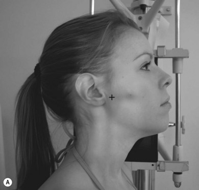
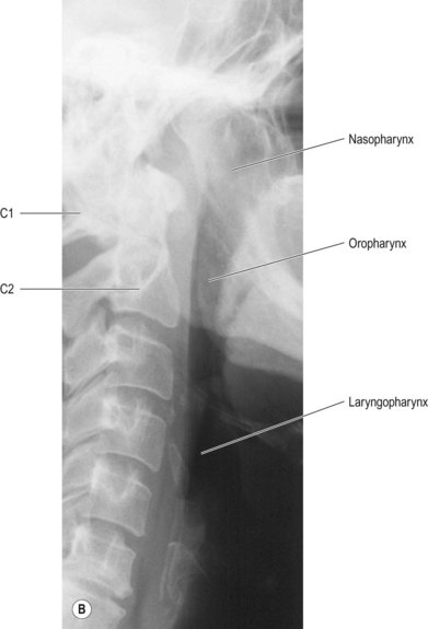
For all projections of the sinuses and postnasal space the image receptor (IR) is vertical
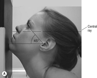
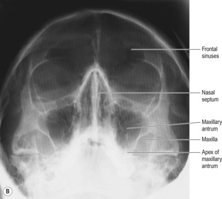
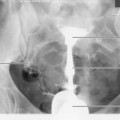
Occipitomental (OM) sinuses (Fig. 19.1A,B)
Positioning
• The patient is seated, facing the IR
• The chin is placed in contact with the midline of the IR and the chin position is adjusted until the orbitomeatal baseline (OMBL) has been raised 45° from the horizontal
• The median sagittal plane (MSP) is perpendicular to the IR, which is assessed by checking that the external auditory meatuses (EAMs) or lateral orbital margins are equidistant from it
Beam direction and focus receptor distance (FRD)
Horizontal, at 90° to the IR and making an angle of 45° with the OMBL
Criteria for assessing image quality
• All paranasal sinuses are demonstrated
• Symmetry of facial bones on each side; equal distance of lateral orbital margins from outer table of temporal bones
• Upper border of the petrous portion of the temporal bone is level with the apex of the maxillary antra
• Images of premolars and molars are medial to, and clear of, the medial aspects of the maxillary sinuses
• Zygomatic arches are seen as a tight ‘C’ and reversed tight ‘C’ laterally
• Sharp image demonstrating the air-filled regions of the paranasal sinuses in contrast with the bones of the skull
| Common errors | Possible reasons |
|---|---|
| Asymmetry of the facial structures | Rotation about MSP |
| Position of the petrous ridge is too high; it is seen through the maxillary sinuses | Chin is not raised enough. (see further notes in Ch. 18, OM facial bones) |
| Petrous ridge is below the maxillary sinuses; image of crowns of premolars overlying the medial aspects of the maxillary sinuses. The frontal sinuses are foreshortened and may appear over-dark | Chin is raised too high |
| or Position appears acceptable; frontal sinuses are over-darkened | Collimation might not be tight enough around the area of interest, thus scatter may blacken the upper anterior aspect of the frontal bone |
Stay updated, free articles. Join our Telegram channel

Full access? Get Clinical Tree


