Chapter 126
Parathyroid Adenoma
Epidemiology
Parathyroid adenoma is the most common cause of primary hyperparathyroidism, accounting for almost 80 to 85% of the cases. Parathyroid adenoma is generally a solitary lesion and affects a single parathyroid gland, however multiple adenomas may be seen in 2 to 3% of cases. They show no sex predilection and may occur at any age, although the middle-aged adults are most commonly affected. The typical sites for a parathyroid adenoma are behind the thyroid gland, behind the strap muscles, or 1 to 2 cm away from the lower pole of the thyroid within the thyrothymic ligament. Infrathyroid lesions are generally spherical. Intrathyroid parathyroid adenomas occur in 1 to 2% of the cases.
Clinical Features
Most patients are asymptomatic. Symptoms include fatigue, weakness, and mental disturbances. Patients with significant hypercalcemia may present with confusion, lethargy, and hyporeflexia.
Pathology
Grossly, parathyroid adenomas are well-encapsulated, soft, yellowish tumors. On histological examination, they are composed principally of chief cells, but many transitional and oxyphil cells are often present. The cells are arranged in large islands or broad bands. A rim of normal or atrophic parathyroid tissue with scattered fat cells can be seen external to the capsule, helping to differentiate an adenoma from diffuse hyperplasia.
Treatment
Surgical parathyroidectomy is the only definitive treatment for primary hyperparathyroidism. Oral phosphates may be given to lower serum calcium temporarily.
Imaging Findings
US
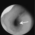
Parathyroid adenomas are usually hypoechoic and well-defined masses with a very sharp edge between the adenoma and the adjacent thyroid parenchyma.
Nuclear Medicine Study