78 Partial Fourier
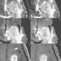
Figure 78.1 presents (A) full and (B) half-Fourier postcontrast T1-weighted spin echo scans, respectively, in a patient with a ring enhancing metastasis (arrow, A) from non-small cell lung carcinoma. One scan average or acquisition was performed for (A), whereas partial Fourier imaging was implemented for (B) (specifically a four-eighths acquisition). The image in (B) was acquired in just over half the scan time of that of (A). The reduction in scan time is due to the implementation of partial Fourier imaging, with fewer phase-encoding steps acquired (in clinical practice, typically four-eighths to seven-eighths). The data as acquired in k space for (A) is presented in Fig. 78.1.C and that for (B) in Fig. 78.1D. Note that just over half of k space is sampled in (D). Spatial resolution is preserved with partial Fourier imaging and is thus identical for (A) and (B). However, there is a resultant decrease in signal-to-noise ratio (SNR) due to the implementation of partial Fourier imaging, reflected by the subtle increase in graininess of white matter seen in (B).
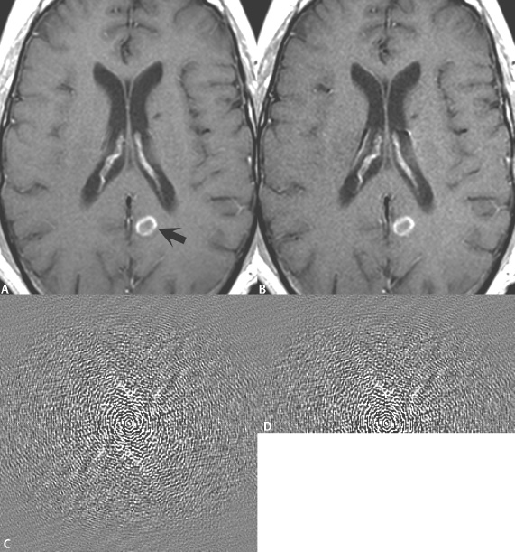
Fig. 78.1
Stay updated, free articles. Join our Telegram channel

Full access? Get Clinical Tree


