Hepatocellular carcinoma (HCC)
Patterns of tumour spread
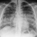
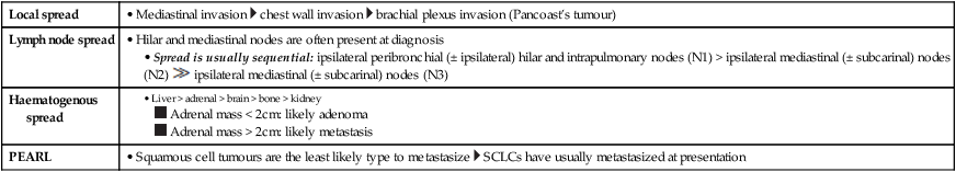
INITIAL COMMON PATTERNS OF TUMOUR SPREAD
Local spread
• Mediastinal invasion  chest wall invasion
  brachial plexus invasion (Pancoast’s tumour)
Lymph node spread
• Hilar and mediastinal nodes are often present at diagnosis
• Spread is usually sequential: ipsilateral peribronchial (± ipsilateral) hilar and intrapulmonary nodes (N1) > ipsilateral mediastinal (± subcarinal) nodes (N2)  ipsilateral mediastinal (± subcarinal) nodes (N3)
Haematogenous spread
PEARL
• Squamous cell tumours are the least likely type to metastasize  SCLCs have usually metastasized at presentation
Local spread
• Local spread into adjacent structures (e.g. pancreas, colon, spleen)
Lymph node spread
• Perigastric: pericardial  lesser curvature
  greater curvature
  suprapyloric
• Extraperigastric: left gastric  common hepatic
  coeliac
  splenic hilum and artery
  hepatic pedicle
  retropancreatic
  mesenteric root
  middle colic
  para-aortic
• NB: retropancreatic, para-aortic and mesenteric nodes are classified as M1 metastatic disease
Haematogenous spread
• Via the portal vein to the liver (25% at presentation)  a similar number will have peritoneal metastases
PEARL
• Transcoelomic spread can occur through the peritoneum (e.g. Kruckenberg tumours)
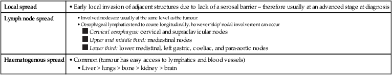
Local spread
• Early local invasion of adjacent structures due to lack of a serosal barrier – therefore usually at an advanced stage at diagnosis
Lymph node spread
Haematogenous spread
• Common (tumour has easy access to lymphatics and blood vessels)
• Liver > lungs > bone > kidney > brain
Local spread
• Invasion through the bowel wall into the perirectal fat – an important predictor of local recurrence and survival
• Extramural venous invasion is an adverse prognostic factor
Lymph node spread
• From the level of the tumour cranially within the mesorectum – proximal blockage (e.g. extensive adenopathy) may cause retrograde spread with lower rectal tumours rarely spreading to the inguinal nodes
• Pelvic side wall spread is unusual
Haematogenous spread
• Liver (via the portal vein)
PEARLS
• Involvement of the circumferential resection margin (CRM) is an adverse prognostic feature (including disruption during surgery)
• Perforation of the peritoneal membrane can result in transcoelomic spread as well as an increased risk of recurrence
• Transcoelomic spread favours the lower right small bowel mesentery and the pouch of Douglas
Local spread
• Vascular invasion of the portal or hepatic veins
Lymph node spread
• Spread to lymph nodes along the hepatoduodenal ligament
Haematogenous spread
• Lung = bone > adrenals > peritoneum
PEARL
• Abdominal lymph nodes are often enlarged with cirrhosis
Local spread
• 70% of tumours arise within the pancreatic head
• Tumour spreads by direct perivascular and perineural invasion
• Head/uncinate process tumours: these usually extend along the SMA and mesenteric root
• Body/tail tumours: these usually infiltrate the coeliac, hepatic or splenic arteries
• Local invasion can involve the stomach, duodenum and retroperitoneum
Lymph node spread
• Early micrometastases at presentation are common
• Primary drainage: superior, inferior, anterior, posterior and splenic lymph nodes
• Secondary drainage: porta hepatis, common hepatic, coeliac, mesenteric root lymph nodes
• Tertiary drainage: peri-aortic and distal superior mesenteric lymph nodes
Haematogenous spread
• Early micrometastases at presentation are common
• These usually involve the liver and peritoneal surfaces
PEARL
• Usually only tumours of the head and uncinate process are surgically resectable (tumours of the body and tail usually have perivascular or perineural metastases at presentation)
Local spread
• Vascular invasion of the portal or hepatic veins
Lymph node spread
• Spread to lymph nodes along the hepatoduodenal ligament – with a propensity to spread to the portocaval lymph node chain as well as the anterior and posterior pancreaticoduodenal chain
Haematogenous spread
• Less common than with HCC – remote metastases are usually to the lung (less commonly to bone, the adrenals and peritoneum)
Local spread
• Perinephric fat  ipsilateral adrenal
  adjacent viscera (including muscles)
• Renal vein invasion (± IVC)
Lymph node spread
• Via lymphatics following the renal vessels to the ipsilateral para-aortic nodes  direct connections with the thoracic duct and mediastinum also exist
Haematogenous spread
• Common sites: lungs > bones, CNS, adrenals
PEARL
• IVC tumour thrombus extending above the hepatic veins requires a transthoracic surgical approach – right atrial involvement requires cardiopulmonary bypass
Local spread
• Initially perivesical fat infiltration  subsequent invasion of adjacent pelvic organs and the pelvic side wall by direct invasion
Lymph node spread
Related posts:
![]()
Stay updated, free articles. Join our Telegram channel

Full access? Get Clinical Tree

 Get Clinical Tree app for offline access