KEY WORDS
Cavernosal Artery. Bilateral arteries centrally placed within the corpus cavernosum. Flow in the artery is measured to detect arterial insufficiency.
Corpora Cavernosum. Two paired tubular structures of erectile cavernous tissue in the penis that become filled with blood during an erection.
Corpus Spongiosum. Third tubular structure of erectile cavernous tissue that lies in front (anterior) and between the paired corpora cavernosa. The urethra lies in the center of it.
Flaccid. Relaxed and without muscle tone.
Fossa Navicularis. Focally dilated portion of the urethra within the glans penis.
Glans Penis. Head of the penis.
Impotence. The inability of the male patient to achieve or maintain erection.
Peyronie’s Disease. A painful curvature of the penis during erection due to fibrous plaques.
Papaverine. A substance that causes an erection by dilating blood vessels when injected directly into the penis.
Priapism. Abnormal persistent erection of the penis, accompanied by pain and tenderness.
Prostaglandin. A hormone that causes an erection when injected directly into the penis.
Sildenafil, Tadalafil, and Vardenafil. Enzyme inhibitors that may be taken orally to cause an erection (“Viagra”).
Sonourethrography. Ultrasound of the urethra while injecting fluid into the urethra.
Stricture. The narrowing of a tube or opening, in this case involving the urethra.
Tunica Albuginea. A fibrous coat around the penis that surrounds the corpora cavernosa.
Urethra. The tubular canal that extends from the bladder to the tip of the penis, through which urine passes.
Venous Incompetence. When referring to a failed erection, a problem caused when blood leaks out of the penis faster than normal, thereby leading to an inability to sustain an erection.
The Clinical Problem
The penis is easy to examine with ultrasound because its internal anatomy is superficial, thereby permitting use of a high-resolution, high-frequency transducer. Four main clinical problems are encountered for which sonography may be of help:
1. Peyronie’s disease. Calcified or fibrous tissue is deposited in the dorsal (Fig. 27-1) portion of the penis so that the organ deviates and is painful when it is erect. The extent of the disease may be defined by ultrasound.
2. Stricture. The length of a stricture and the width of the stricture walls may be determined when the urethra is distended with fluid (the patient’s urine, iatrogenically introduced saline, or gel) while imaging with a linear array transducer over the relevant area.
3. Impotence due to poor penile arterial flow. Arterial flow can be calculated using pulsed Doppler studies. An inadequate systolic flow indicates arterial insufficiency.
4. Impotence due to venous leak. An inadequate erection occurs because venous blood “leaks” out during attempted erection.
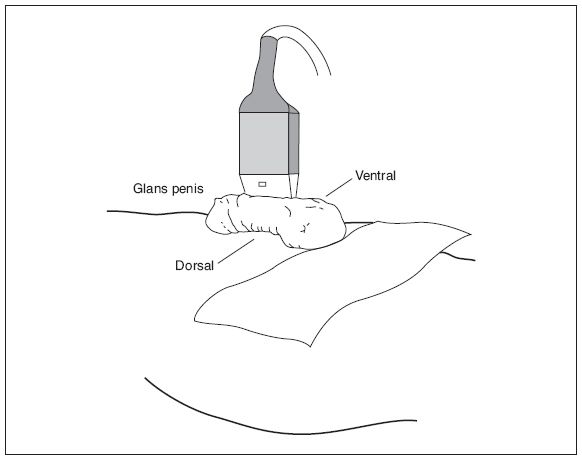
Figure 27-1. ![]() Position used for examining the penis in penile flow studies. It is also a good position for evaluating the penis for Peyronie’s disease. The penis is in the anatomic position.
Position used for examining the penis in penile flow studies. It is also a good position for evaluating the penis for Peyronie’s disease. The penis is in the anatomic position.
Anatomy
The penis is considered in correct anatomic position when the dorsum lies against the abdomen, exposing the ventral side (Fig. 27-1). The penile portion of the urethra is midline, ventral, and surrounded by the corpus spongiosum. Posterior and lateral to the urethra are two vascular structures called the corpora cavernosa. All three components are surrounded by fibrous tissue called the tunica albuginea. Contained within each corpora cavernosum is erectile tissue and a cavernosal artery. In the dorsal portion of the penis are the deep dorsal artery and vein, and the superficial dorsal vein (Fig. 27-2).
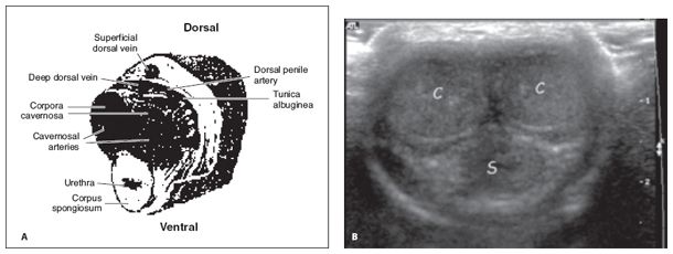
Figure 27-2. ![]() A. View showing the position of the cavernosal arteries within the corpora cavernosa and of the urethra within the corpus spongiosum. Note the tunica albuginea surrounding the corpora. B. Corresponding transverse sonogram showing the paired corpora cavernosa (C) and corpus spongiosum (S).
A. View showing the position of the cavernosal arteries within the corpora cavernosa and of the urethra within the corpus spongiosum. Note the tunica albuginea surrounding the corpora. B. Corresponding transverse sonogram showing the paired corpora cavernosa (C) and corpus spongiosum (S).
Technique
Related posts:
Stay updated, free articles. Join our Telegram channel

Full access? Get Clinical Tree


