Fig. 44.1
Establishing access window. (a) Straight clamp is placed along the sonographically determined liver margin (LM) and palpated right rib margin (RM). Barium given the night before is present within the transverse colon (TC). Insufflation of the stomach demonstrates an accessible window to the gastric antrum (GA). (b) Four 25-gauge needles are placed at the window location and demarcate the future location of the T-tacks (Image (b) courtesy of Diego Covarrubias MD)
If the patient has not had barium the night before, the interventional radiologist may first look with fluoroscopy to see if there is a fortuitous gas-filled colon. In more ambiguous cases, air or water soluble contrast enema should be administered to demarcate the colon. If a nasogastric tube has not been placed, often in the outpatient setting or in patient with history of head/neck cancer, this may be done under fluoroscopic guidance, typically using a 5-Fr Kumpe catheter with a 0.035 3-J wire to maneuver it into the stomach.
44.1.3.2 Imaging Modality
PRG is typically performed under fluoroscopic guidance and rarely under CT guidance. CT-guided placement may be used in situations where the window between the liver, transverse colon, and ribs is too small or nonexistent, often requiring a different trajectory. CT may also be used in situations where the stomach cannot be insufflated via nasogastric tube (e.g., esophageal stricture precluding nasogastric tube placement or gastrostomy of a gastric remnant of patients status post-Roux-en-Y gastric bypass); however, a recently published technique of modified radiology-guided percutaneous placement using contrast injection via a 21-gauge needle to localize the stomach and then fill it with air has provided a successful fluoroscopic approach in circumstances of complete obstruction of the upper digestive tract (Chan et al. 2011).
44.1.3.3 Equipment
There are two basic types of catheters that may be placed for a gastrostomy. One type is a low-profile (button) catheter (Fig. 44.2a) to which tubing may be connected for feeding and which is secured in the stomach with either a balloon or mushroom tip. The other type is a Cope catheter (Fig. 44.2b) that has an external segment and locking loop pigtail to prevent it from exiting the stomach. Both types use gastropexy fasteners prior to catheter placement. Choosing the tube type often depends on the physician and patient. Some radiologists prefer to start with the longer tube and possibly switch to one with a lower profile after a mature tract has formed, though placement of button catheters may be done first without the need for tract maturation (Thornton et al. 2002). More active patients may prefer the low-profile tube. Bathing, patient transfer, and dressing are all reported as easier for the elderly with the low-profile tube (Borge et al. 1995).
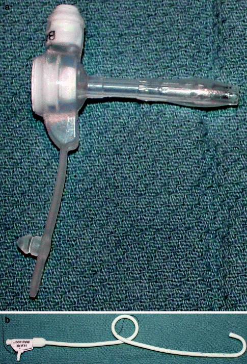

Fig. 44.2
Gastrostomy catheters. (a) Button-type low-profile catheter which hubs at the skin surface with the distal end held within the stomach by an inflatable balloon at the tip. (b) Cope G-tube catheter. The pigtail in the mid portion of the tube keeps the catheter from exiting the stomach lumen via the gastrostomy and may be locked into position by pulling and securing the proximal aspect of the string
44.1.3.4 Gastropexy
Gastropexy is performed prior to catheter placement. Fixation of the stomach to the anterior abdominal wall is thought to help adhesions form and minimize leakage from the stomach, preventing peritonitis and inadvertent migration of the catheter, and allowing for easier tract access should the tube fall out (Thornton et al. 2002; Dewald et al. 1999). There are many different mechanisms for placing the fastener devices. They are typically deployed using a needle tip which houses the fastener; the fastener is deployed after the needle is confirmed to be within the stomach via aspiration of air or injection of contrast (Fig. 44.3a). The internal component of the fastener apposes the gastric mucosa and the external component, either a spongy or plastic pledget, apposes the skin. Ideally, four fasteners are placed; however, sometimes anatomic constraints only allow three.
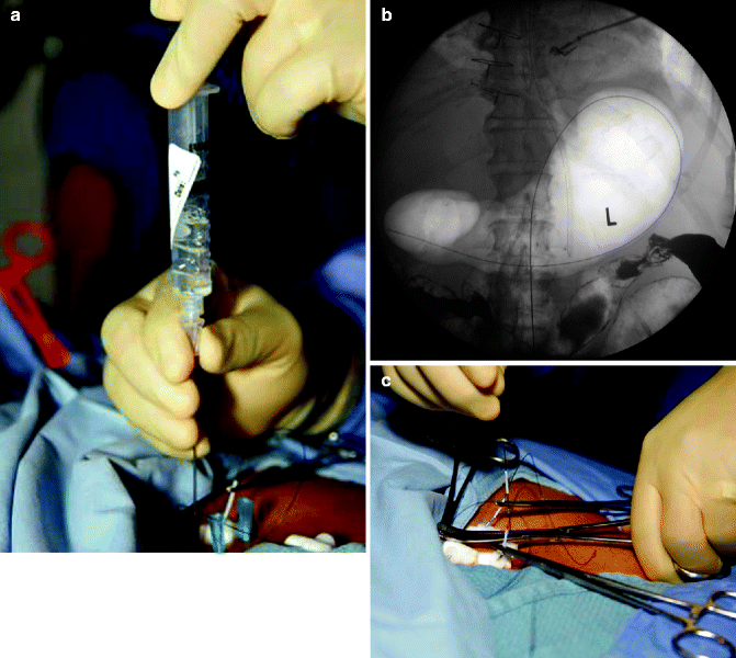

Fig. 44.3
Gastropexy. (a) T-fasteners are placed at sites demarcated by 25-gauge needles. A hollow bore needle containing the fastener is advanced into the gas-filled stomach and air is aspirated to confirm entry. (b) Wire is placed into the needle deploying the fastener and confirming intraluminal location. (c) Fasteners are secured into place after all four are deployed, thereby pexying the stomach to the anterior abdominal wall. (Images (a, c) courtesy of Diego Covarrubias M.)
After placing the fasteners, the stomach is pexied to the anterior abdominal wall by cinching them (Fig. 44.3c). Care must be taken to achieve a balance between cinching the fasteners too loose and thereby serving no purpose and too tight and potentially inducing pressure necrosis.
44.1.3.5 Catheter Placement
When placing a Cope catheter, via an incision central to the fasteners, a wire is placed into the stomach and over this wire, the tract is dilated until it can accommodate a peel-away sheath which eases passage of the blunt end gastrostomy tube down the tract and into the stomach (Fig. 44.4a). If PRGJ is required, then the wire should be directed past the pylorus and looped within the small bowel lumen to the jejunum (Fig. 44.4c). Often torqueing of the dilator/peel-away sheath combination is helpful in directing the wire, or a Kumpe catheter may also be used to this end. The gastrostomy or gastrojejunostomy tube is placed over the wire and secured using the looping–locking mechanism of the Cope catheter (Fig. 44.4b). Contrast should be injected via the catheter to ensure that the tube is positioned appropriately (Fig. 44.4b, d).
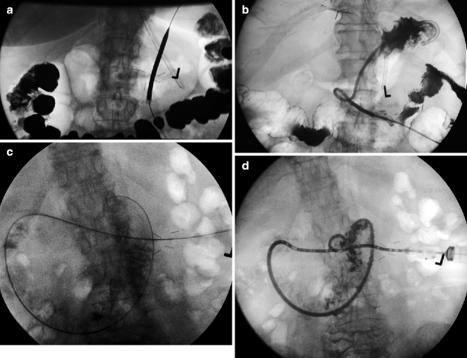

Fig. 44.4
PRG and PRGJ catheter placement. (a) Peel-away sheath is placed central to the fasteners that secure the stomach. (b) The blunt-tipped gastrostomy tube is placed over a wire and through the peel-away sheath, with contrast injection confirming location. (c) Wire is passed beyond the ligament of Treitz in preparation for gastrojejunostomy placement. (d) After placement of the peel-away sheath (not shown), the gastrojejunostomy tube is placed over the wire to the jejunum. Contrast injection confirms the location
When placing a button tube, the length of the tract from the skin to the stomach must first be measured using the device in the kit which consists of a terminal balloon and is marked with calibrations on its length. After the tract length has been determined, the appropriate length and French catheter should be chosen and placed down the tract and the tip balloon should be filled to the specified volume.
44.1.4 Catheter Care
Typically, gastrostomy catheter usage ensues 15–24 h after placement; the wait helps to decrease the chance of leakage from the newly formed hole within the stomach. Jejunal feedings may be started 0–4 h post-procedure (Hicks et al. 1990).
The external string of the fasteners of the gastropexy is cut after about 2 weeks, by which time adhesions should have formed, securing the stomach to the anterior abdominal wall. Cutting the external string allows the inner securing components of the fasteners to fall into the stomach and be excreted.
Rigorous skin care including keeping the skin site clean and dry and vigilance against any evidence of pressure necrosis while the fasteners are still in place is mandatory. Catheters that become dislodged or pulled out may often be rescued if enough time has passed for a tract to form and the rescue is done a timely manner. If there will be a delay in replacing the tube, a small French catheter (e.g., pediatric feeding tube) may be placed down the tract to maintain its patency. Clogged or kinked tubes may need to be replaced over a wire using fluoroscopic guidance.
44.1.5 Conclusion
PRG and PRGJ is an effective procedure with low morbidity and mortality and provides enteral nutrition to those unable, either permanently or temporarily, to eat or as a method of gastric drainage in the chronically obstructed patient. The procedure serves as a good replacement for a nasogastric tube when tube dependence is expected to exceed a few weeks. Furthermore, given the superior safety profile of radiologic placement of gastrostomy and gastrojejunostomy tubes compared with surgically and endoscopically placed tubes, PRG/PRGJ should be considered in the elderly.
44.2 Splanchnic Neurolysis
44.2.1 Introduction
The celiac plexus is a network of nerves located in the retroperitoneum flanking the anterolateral aorta near the celiac, superior mesenteric, and renal artery branches. The plexus includes the gastric, pancreatic, splenic, hepatic, and suprarenal plexuses and innervates the liver, gallbladder, biliary tract, pancreas, spleen, adrenal glands, kidneys, mesentery, and small and large bowel, proximal to the transverse colon. As a relay center for pain impulses of the upper abdomen, lysis of the plexus serves as a method to treat refractory pain requiring significant opioid use.
Stay updated, free articles. Join our Telegram channel

Full access? Get Clinical Tree


