Abstract
The differential of peritoneal lesions includes both malignant (metastases, lymphoma, primary peritoneal malignancies) and benign (bowel perforations, pelvic inflammatory disease) etiologies. Peritoneal carcinomatosis may be visible on both FDG PET and CT, appreciated on FDG PET only, appreciated on CT only, or be occult on both FDG PET and CT and identified during surgerical exploration.
Keywords
FDG, PET/CT, peritoneum, carcinomatosis, mesothelioma
The peritoneum is a membranous lining of the abdominal cavity and organs. The parietal peritoneum lines the abdominal cavity, whereas the visceral peritoneum lines the intraperitoneal organs. Between layers of the peritoneum is the peritoneal cavity, a potential space. This potential space is normally not identifiable on imaging studies, unless the space becomes filled with fluids, air, or masses. Masses in the peritoneum include both benign and malignant etiologies. Malignancies involving the peritoneum include metastases, lymphoma, and primary malignancies such as mesothelioma and desmoplastic round cell tumor. Benign peritoneal masses include infections and omental infarcts.
Peritoneal Metastases
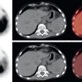
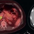
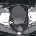
The most common peritoneal malignancy is metastatic disease. The most common primary malignancies to cause peritoneal metastases are colon, ovary, pancreas, and stomach (the “COPS”), although nearly any primary malignancy may result in peritoneal metastases when advanced. Diffuse peritoneal metastases are often referred to as peritoneal carcinomatosis. When peritoneal carcinomatosis is noted and a primary malignancy has not been identified, check the COPS for masses or wall thickening. On computed tomography (CT), peritoneal metastases may present as nodules or thickening of the peritoneal membranes and mesentery or thickening/implants along the surface of the bowel or abdominal organs and may often be accompanied by peritoneal free fluid. Peritoneal metastases may be 18F-fluorodeoxyglucose (FDG) avid and if avid may identify sites of disease not appreciated on anatomic imaging. Overall, peritoneal metastases may be seen on both FDG positron emission tomography (PET) and CT ( Fig. 17.1 ), FDG PET only ( Fig. 17.2 ), or CT only ( Fig. 17.3 ) and unfortunately may be present even when not identified on either CT or FDG PET. Diffuse peritoneal carcinomatosis may be occult on all known imaging modalities but then identified during surgical exploration.


There are multiple peritoneal reflections which invaginate into the abdominal organs. Examples include the falciform ligament and fissure for the ligamentum venosum along the surface of the liver, as well as the splenic hilum. If possible, differentiate between peritoneal metastases on these peritoneal invaginations from parenchymal organ metastases ( Fig. 17.4 ). Often, parenchymal organ metastases have a different clinical significance from peritoneal metastases. An unusual peritoneal metastasis is the Sister Mary Joseph “node” ( Fig. 17.5 ). This is a peritoneal metastasis that protrudes out of the umbilicus. Sister Mary Joseph was an assistant at St. Mary’s Hospital in Rochester, Minnesota, who noticed that patients with abdominal malignancies who developed a nodule at the umbilicus had very poor prognoses. The term was subsequently eponymously named for Sister Mary Joseph.