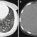
Fig. 18.1
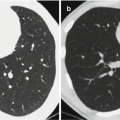
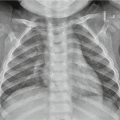
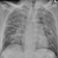
Pertussis complicated by pneumonia. CT scanning demonstrates thickened vascular bundle of bronchus of both lungs, with poorly defined boundaries and flakes of ground-glass opacities and patches of shadows (Reprint with permission from Abe and Watanabe Pediatr Emerg Care, 2003, 19(4): 262)
18.7.2 Central Nervous System
The encephalopathies complicating pertussis include encephaledema and cerebral hypoxia that commonly involve the nuclei in basal ganglia.
18.7.2.1 CT Scanning
Encephaledema and cerebral hypoxia commonly occur in basal ganglia, which is demonstrated as symmetric low-density shadows or scattering low-density shadows with poorly defined boundaries. There are also demonstrations of blurry interface between gray and white matter and absence of some sulci. Brain parenchymal hemorrhage is demonstrated as spots, patches, and round shadows with high density in the brain parenchyma, which are possibly surrounded by low-density edema zone in different widths. Subarachnoid hemorrhage can be demonstrated as the absence sulci and cistern as well as increased density in cast-like appearance.
18.7.2.2 MR Imaging
Acute encephaledema and cerebral hypoxia commonly occur in the basal ganglia, which are demonstrated by symmetric long T1 long T2 signals. Otherwise, they can be demonstrated as multifocal or diffusive flakes of long T1 long T2 signals. By DWI, cytotoxic cerebral edema is demonstrated as high signal with obviously decreased ADC value, and interstitial cerebral edema is demonstrated as no high signals and slightly or moderately increased ADC value. In cases with acute hematoma, MR imaging demonstrates equal signal by T1WI and slightly decreased signal by T2WI. In cases with subacute and chronic hematoma, MR imaging demonstrates high signals by both T1WI and T2WI (Fig. 18.2).
Case Study 2
A boy aged 6 years, he had a history of vaccination against DPT. By physical examination, T was 36.3 °C and WBC 12 × 109/L.
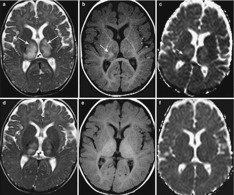

Fig. 18.2
Pertussis complicated by encephalopathy. (a) At day 3 after admission, MR imaging demonstrates symmetrical increased signal in bilateral basal ganglia and posterior limb of internal capsule by T2WI (pointed by arrows). (b) T1WI demonstrates high signal in the right thalamus, indicating cerebral hemorrhage (pointed by arrow). (c) Increased ADC value in bilateral thalamus and internal capsule, decreased ADC value in the high-signal area of the right thalamus by T1WI, indicating cerebral hemorrhage or cytotoxic cerebral edema (pointed by arrow). (d) Reexamination after 1 week, T2WI demonstrates decreased signal and range of the lesions in the bilateral thalamus. (e) T1WI demonstrates no increase of signal intensity of the lesions in the right thalamus. (f) Absence of the area with increased ADC value in the right thalamus (Reprint with permission from Aydin H, et al. Pediatr Radiol, 2010, 40(7): 1281)
18.7.3 Fracture
Fracture rarely occurs in the cases of pertussis. In the cases with pertussis complicated by fracture, X-ray and CT scanning demonstrate continual broken bone, with favorable demonstration of the location and quantity of fracture as well as displacement of fracture.
Case Study 3
A boy aged 11 years complained of sudden right chest pain for 3 h. He was diagnosed with pertussis 6 weeks ago.
For case detail and figures, please refer to Prasad and Baur, J Paediatr Child Health, 2001, 7(1): 91.
Stay updated, free articles. Join our Telegram channel

Full access? Get Clinical Tree


