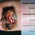
Fig. 6.1 Patient with newly diagnosed neck left melanoma. Lymphoscintigraphy following subdermal injection of 99mTc-antimony colloid was performed. Surface-rendered SPECT/CT (a) demonstrates high uptake at the injection site (red arrow) with activity flare evident on axial SPECT/CT (b) owing to the high number of counts. A left lower cervical sentinel node was identified (c). Wide local excision and sentinel node biopsy was performed, which was negative. Twelve months later, the patient presented with a left neck lump. FDG PET/CT demonstrated recurrence in a level II node deep the site of prior WLE (d, e, f). The location of recurrence was deep in the site of injection on the SLN SPECT/CT study and likely masked by high intensity of activity from the overlying site of injection
Teaching points:

SPECT/CT enables precise localisation of sentinel nodes, but there is reduced sensitivity near the site of injection owing to flare of activity which may result in false-negative findings.
Fig. 6.2 Patient with mid-back thick melanoma scheduled for sentinel node biopsy. Four injections of 99mTc-antimony colloid were administered by subdermal injection on either side of the scar. Surface-rendered SPECT/CT images (a) and (b) demonstrate high uptake at the injection site and three separate pathways of drainage, to the left prescapular region (a), right axillary region (b) and an atypical location in the left lower back. Posterior to the left paraspinal muscles (c), corresponding to a tiny lymph node on CT (d). Further interrogation of SPECT/CT images demonstrates extension of activity from this location to the retroperitoneum (red arrow)
Teaching points:

SPECT/CT is able to demonstrate atypical drainage pathways that are difficult to identify on conventional planar imaging.
Drainage from the skin of the back to retroperitoneal and paravertebral lymph nodes is uncommon but described in 2.5% of patients with primary back melanomas [1].
Identification of multiple sentinel nodes may present challenges for the surgical team as targeting all sites may not be feasible or result in additional morbidity. Close communication is needed between the nuclear medicine specialists and surgical team in how to deal with such scenarios.
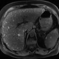
Fig. 6.3 A patient with metastatic melanoma had a 6 mm pulmonary nodule identified on surveillance CT (a). FDG PET/CT demonstrated focal intense uptake in this nodule suggesting metastatic disease, low dose CT (top) and fused PET/CT (bottom). The patient underwent excision via video-assisted thoracoscopic surgery (VATS) with histopathology confirming the diagnosis of metastatic melanoma. Follow-up CT performed 6 months (b) later demonstrated a new soft tissue abnormality within the surgical bed suggesting local recurrence. FDG PET/CT, however, demonstrated no metabolic activity in the area of concern and the finding was interpreted as benign postsurgical change. Subsequent follow-up demonstrated stable findings confirming benign aetiology
Teaching points:


Although sensitivity of PET diminishes with sub-cm lesions, especially in the lung which is subject to respiratory motion, metastatic melanoma is usually highly metabolically active facilitating characterisation of small abnormalities.
The sensitivity of PET has also improved significantly with modern generation devices incorporating improvements including time-of-flight, point-spread-function modelling, improved attenuation correction, reconstruction algorithms and respiratory gating.
PET is able to differentiate postsurgical changes from recurrent disease.
Fig. 6.4 A middle-aged lady presented with left groin nodal recurrence on the background of prior left leg primary. FDG PET/CT (a) prior to planned lymphadenectomy demonstrated additional sub-cm external iliac nodal metastases and a posterior mediastinal nodal metastasis (b). The patient proceeded to extended lymphadenectomy followed by radiotherapy to mediastinal nodal disease; radiotherapy treatment plan shown in (c). Restaging FDG PET/CT 4 months after completion of radiotherapy demonstrated a complete metabolic response in the posterior mediastinum (d) but new focal intense uptake in the right atrium (e). In correlation with the radiation treatment plan and annular morphology which is best appreciated on the maximum intensity projection (MIP) image (f), this was interpreted as post-radiotherapy change within the segment of irradiated myocardium. Follow-up PET (not shown) demonstrated partial resolution of this finding with this time
Related posts:


Stay updated, free articles. Join our Telegram channel
Full access? Get Clinical Tree
