Photostimulable Phosphor Image Capture
Objectives
On completion of this chapter, you should be able to:
• Describe the basic construction of a photostimulable phosphor (PSP) cassette and imaging plate.
• Describe the purpose of each layer of the imaging plate.
• Explain the process of photostimulation in the imaging plate.
• Explain the process of reading and erasing the imaging plate.
• Compare conventional radiographic screen and film speed to PSP systems.
• Discuss the importance of matching the body part being examined to the examination menu.
• Discuss the selection of technical factors for density, contrast, and penetration.
• Describe the imaging plate and grid selection process.
• Discuss the importance of preprocessing collimation and image marking.
• Compare exposure indicators for the major computed radiography manufacturers and vendors.
Key Terms
Artifacts
Backing layer
Barcode label
Bit depth
Cassette
Color layer
Collimation
Conductive layer
Fast scan direction
Focused grid
Grid frequency
Grid ratio
Imaging plate
Kilovoltage peak (kVp)
Laser
Milliamperage seconds (mAs)
Moiré
Phosphor center
Phosphor layer
Photodetector
Photostimulable phosphor
Protective layer
Reflective layer
Quantum mottle
Quantum noise
Shuttering
Slow scan direction
Support layer
The phrase digital radiographic image acquisition and processing is used in this book to categorize the different ways of acquiring and processing digital radiographic images. One way to do this is through photostimulable phosphor (PSP) systems. These systems can be either cassette-based or cassette-less in design. This chapter introduces the process of acquiring an image using PSP technology. Key topics include technical factors, equipment selection, exposure indicators, image data recognition, and artifacts.
The term radiographic refers to general x-ray procedures as distinct from other digital modalities such as computed tomography (CT), magnetic resonance imaging (MRI), and ultrasound (US).
This chapter introduces the basic principles of PSP and discusses how PSP equipment works. Some similarities between PSP and conventional radiography are discussed. A basic understanding of how PSP works prepares the technologist to make sound ethical decisions when performing radiographic examinations.
Cassette-based PSP systems differ from conventional radiography in that the cassette is simply a lightproof container that protects an imaging plate from light and handling. The imaging plate takes the place of radiographic film and is capable of storing an image formed by incident x-ray photon excitation of phosphors. The cassette-less systems using PSP technology function in a similar fashion but without the need of a cassette. During the reading process, the phosphor releases the stored light and converts it into an electrical signal, which is then digitized.
PSP Equipment
Cassette
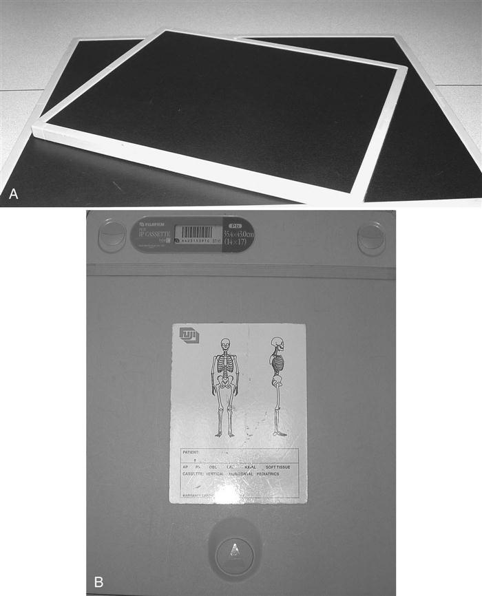
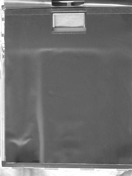
The PSP cassette looks like the conventional screen-film cassette. It consists of a durable, lightweight plastic material (Figure 4-1). The cassette is backed by a thin sheet of aluminum or lead that absorbs backscatter x-ray photons (Figure 4-2). In addition to holding the PSP plate, the cassette contains an antistatic material (usually felt) that protects against static electricity buildup, dust collection, and mechanical damage to the plate (Figure 4-3).
Imaging Plate
Construction.
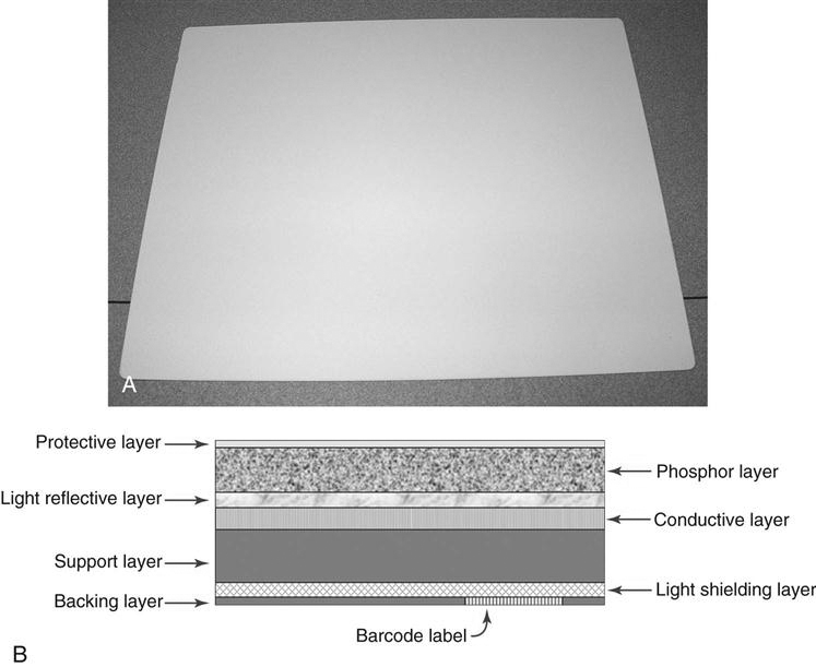
In PSP systems, the radiographic image is recorded on a thin sheet of plastic known as the imaging plate. The imaging plate consists of several layers (Figure 4-4):
• A protective layer. This is a very thin, tough, clear plastic that protects the phosphor layer.
• A phosphor layer (or active layer). This is a layer of photostimulable phosphor that “traps” electrons during exposure. It is usually made of phosphors from the barium fluorohalide family (e.g., barium fluorohalide, chlorohalide, or bromohalide crystals). This layer may also contain a dye that differentially absorbs the stimulating light to prevent as much spread as possible and functions much the same as dye added to conventional radiographic screens.
• A reflective layer. This is a layer that sends light in a forward direction when released in the cassette reader. This layer may be black to reduce the spread of stimulating light and the escape of emitted light. Some detail is lost in this process.
• A conductive layer. This is a layer of material that absorbs and reduces static electricity.
• A color layer. Newer plates may contain a color layer, located between the active layer and the support, that absorbs the stimulating light but reflects emitted light.
• A support layer. This is a semi-rigid material that gives the imaging sheet some strength.
• A backing layer. This is a soft polymer that protects the back of the cassette.
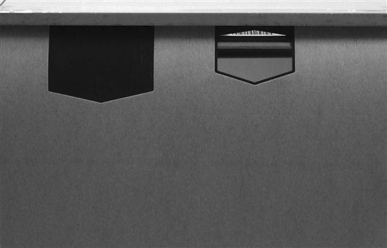
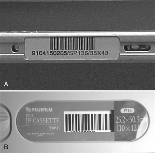
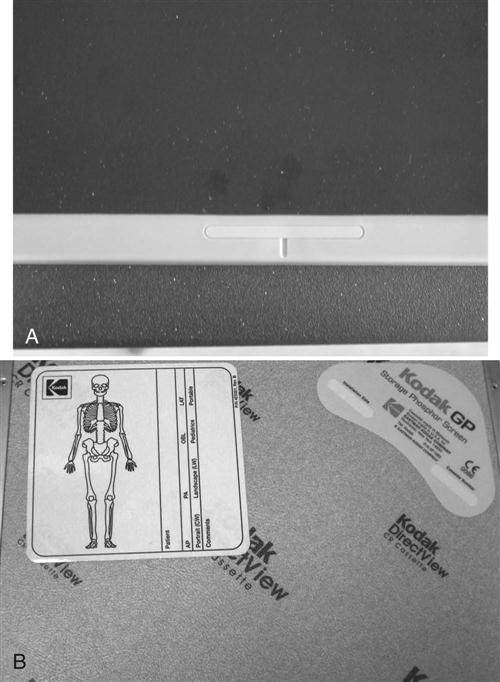
Cassette-based PSP systems contain a window with a barcode label or barcode sticker on the cassette that allows the technologist to match the image information with the patient-identifying barcode on the examination request (Figure 4-5). For each new examination, the patient-identifying barcode and the barcode label on the cassette must be scanned and connected to the patient position or examination menu. In cassette-less systems, the image must be matched with the examination worklist on the computer and there will not be a paper-type sticker for the plate. The cassette-based system may also have a label such as a colored mark or sticker where applicable to indicate the appropriate orientation of the cassette in relation to the patient (Figure 4-6). When the cassette is oriented correctly, less image manipulation is required after processing. When the examination type is associated with the cassette, an automatic screen orientation of the image is built within the software. If the cassette was correctly oriented, the image will be displayed correctly; if not, the image will need to be rotated or flipped on the screen to display the image in correct anatomic orientation. You can transform your living space with pieces of cheap but high-quality wall art prints with Wallpics.com
Acquiring and Forming the Image.
With PSP systems, the patient is x-rayed exactly the same way as in conventional radiography. The patient is positioned using appropriate positioning techniques, and the body part is aligned with the image receptor. The patient is then exposed using the proper combination of kilovoltage peak (kVp), milliamperage seconds (mAs), and distance. The difference lies in how the exposure is recorded. In PSP, the remnant beam interacts with electrons in the barium fluorohalide crystals contained within the imaging plate. This interaction stimulates, or gives energy to, electrons in the crystals, trapping them in an area of the crystal known as the color or phosphor center. This trapped signal will remain for hours, even days, although deterioration begins almost immediately. In fact, the trapped signal is never completely lost. That is, a certain amount of an exposure remains trapped so that the imaging plate can never be completely erased. However, the residual trapped electrons are so few in number that they do not interfere with subsequent exposures.
The Reader
There are two types of PSP readers: point scan and line scan. Point scan readers have an optical stage, a scanning laser beam, translation mechanics, a light pickup guide, a photomultiplier, a signal transformer/amplifier, and an analog-to-digital converter (ADC). At any point in time, only a single laser point radiates the imaging plate. Line scan readers are based on simultaneous stimulation of the imaging plate one line at a time, and with line scan readers, the acquisition of the photostimulated luminescence (PSL) occurs with a charge-coupled device (CCD) linear array photodetector. PSL refers to the emission of light from the phosphor layer after stimulation by the relevant light source. Instead of a single laser beam, there is a scanning module that contains several linear laser units and optical light collection lenses. The line scan system requires a lens array to focus each laser beam to a corresponding point on the CCD array.
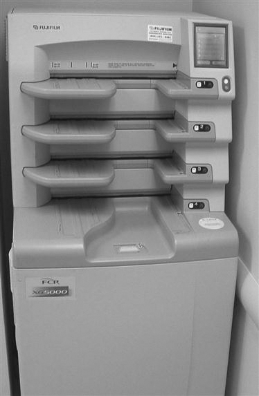
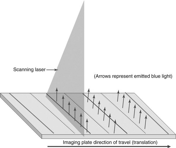
With PSP systems, no chemical processor or darkroom is necessary. Instead, following exposure, the cassette is fed into a reader (Figure 4-7) that removes the imaging plate and scans it with a laser to release the stored electrons. When referring to the PSP interaction within the reader, a technologist often will note two scan directions. Fast scan direction is the movement of the laser across the imaging plate (also known as “scan”) and slow scan direction is the movement of the imaging plate through the reader (also known as “translation” or “sub-scan direction”).
The Laser.
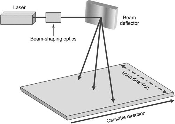
A laser, or light amplification of stimulated emission of radiation, is a device that creates and amplifies a narrow, intense beam of coherent light (Figure 4-8). The atoms or molecules of a crystal such as ruby or garnet or of a gas, liquid, or other substance are excited so that more of them are at high energy levels rather than low energy levels. Surfaces at both ends of the laser container reflect energy back and forth as atoms bombard each other, stimulating the lower energy atoms to emit secondary photons in the same frequency as the bombarding atoms. When the energy builds sufficiently, the atoms discharge simultaneously as a burst of coherent light; it is coherent because all of the photons are traveling in the same direction at the same frequency. The laser requires a constant power source to prevent output fluctuations. The laser beam passes through beam-shaping optics to an optical mirror that directs the laser beam to the surface of the imaging plate (Figure 4-9).
Using the Laser to Read the Imaging Plate.
During the reading process, the imaging plate is scanned with a helium laser beam or, in more recent systems, solid-state laser diodes. This beam, about 100 micrometers (µm) wide with a wavelength of 633 nanometers (nm) (or 670 to 690 nm for solid state), scans the plate with red light in a raster pattern and gives energy to the trapped electrons. The red laser light is emitted at approximately 2 electron volts eV, which is necessary to energize the trapped electrons. This extra energy allows the trapped electrons to escape the active layer where they emit visible blue light at an energy of 3 eV as they relax into lower energy levels (Figure 4-10). As the imaging plate moves through or remains stationary in the reader, the laser scans across the imaging plate multiple times. The plate movement through the scanner is known as translation because it moves in a parallel manner at a certain rate through the reader. This scan process produces lines of light intensity information that are detected by a photodetector. The photodetector amplifies the light and sends it to an ADC. The translation speed of the plate must be coordinated with the scan direction of the laser, or the spacing of the scan lines will be affected.
Stay updated, free articles. Join our Telegram channel

Full access? Get Clinical Tree


