A stream of high energy electrons produced by an electron gun accelerate from a cathode filament and strike a rotating tungsten anode. X-ray photons are generated within the anode which rotates to dissipate heat. The beam of X-ray photons is shielded and coned to reduce the scatter of X-rays produced
1.2 Characteristic radiation generation
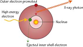
High energy electrons collide with and eject an inner shell tungsten electron (green) with subsequent promotion of an outer shell electron (red) to take its place. X-ray photons of a uniform ‘characteristic’ energy are generated
1.3 Bremsstrahlung radiation
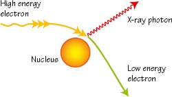
A high energy electron that passes near a tungsten nucleus is deflected and decelerated with generation of an X-ray photon. X-ray photons of variable energy are generated in this way and therefore a non-uniform energy spectrum is produced. This is known as Bremsstrahlung ‘Braking’ radiation
1.4 The X-ray spectrum
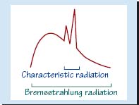
Bremsstrahlung radiation produces a wide spectrum of X-ray energies within the X-ray beam. Characteristic radiation generation however produces a relatively narrow band of X-ray energy. Imaging techniques optimise this characteristic band of X-rays in producing a radiograph
1.5 Image generation
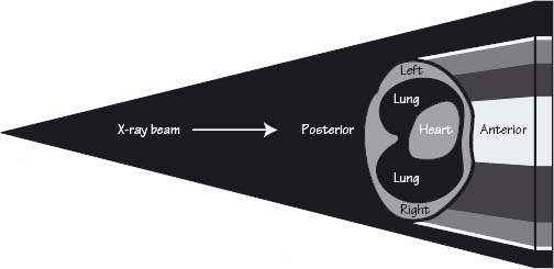
A chest X-ray (CXR) is usually taken with the beam passing from posterior to anterior (PA). The X-ray beam is divergent and so the resultant image is magnified. The closer the patient is to the detector the less magnification is produced. X-rays which hit the detector uninterrupted appear black on the image. Those X-rays that pass into thick structures (e.g. heart) or dense structures (e.g. bones) are attenuated and appear white. Other structures such as the lungs and soft tissues appear as a range of grey, according to their density
Plain XR physics
On 8 November 1895, the German physicist Wilhelm Conrad Röentgen discovered the X-ray, a form of electromagnetic radiation which travels in straight lines at approximately the speed of light. X-rays therefore share the same properties as other forms of electromagnetic radiation and demonstrate characteristics of both waves and particles. X-rays are produced by interactions between accelerated electrons and atoms. When an accelerated electron collides with an atom two outcomes are possible: